Copyright
©2012 Baishideng Publishing Group Co.
World J Gastroenterol. Jul 7, 2012; 18(25): 3327-3330
Published online Jul 7, 2012. doi: 10.3748/wjg.v18.i25.3327
Published online Jul 7, 2012. doi: 10.3748/wjg.v18.i25.3327
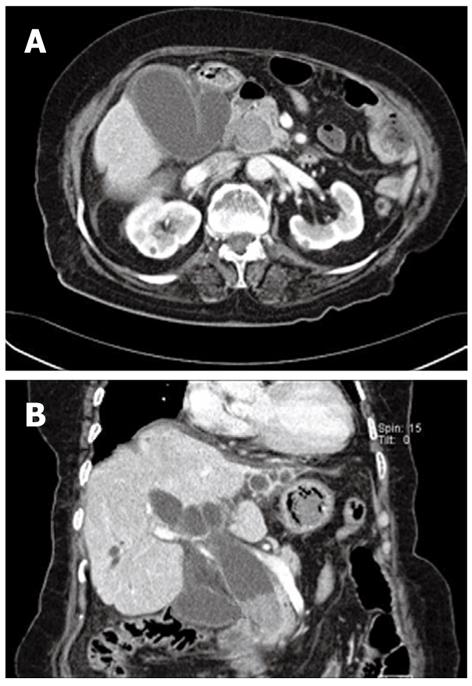
Figure 1 Computed tomogram revealed a huge stone in the distal common bile duct.
Marked dilation of the gallbladder and bile duct was also seen. A: Axial view; B: Coronal view.
- Citation: Chung HJ, Jeong S, Lee DH, Lee JI, Lee JW, Bang BW, Kwon KS, Kim HK, Shin YW, Kim YS. Giant choledocholithiasis treated by mechanical lithotripsy using a gastric bezoar basket. World J Gastroenterol 2012; 18(25): 3327-3330
- URL: https://www.wjgnet.com/1007-9327/full/v18/i25/3327.htm
- DOI: https://dx.doi.org/10.3748/wjg.v18.i25.3327


