Copyright
©2012 Baishideng Publishing Group Co.
World J Gastroenterol. Jun 28, 2012; 18(24): 3177-3180
Published online Jun 28, 2012. doi: 10.3748/wjg.v18.i24.3177
Published online Jun 28, 2012. doi: 10.3748/wjg.v18.i24.3177
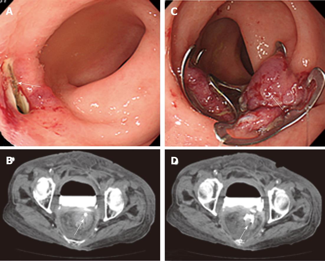
Figure 3 Colonoscopy and computed tomography of case 2.
A: Colonoscopy revealed a large fistula (25 mm) (arrow) in the ventral wall of the rectum; B: Following contrast radiography with a colonoscopy, computed tomography revealed the pooling of the radiocontrast agent in the urinary bladder and fistula (arrow); C: Complete closure of the fistula with a series of over-the-scope-clips (OTSCs) (double OTSCs) was performed (arrow); D: Computed tomography revealed the pooling of the radiocontrast agent in the urinary bladder and the complete closure of the fistula with double OTSCs (arrow).
- Citation: Mori H, Kobara H, Fujihara S, Nishiyama N, Kobayashi M, Masaki T, Izuishi K, Suzuki Y. Rectal perforations and fistulae secondary to a glycerin enema: Closure by over-the-scope-clip. World J Gastroenterol 2012; 18(24): 3177-3180
- URL: https://www.wjgnet.com/1007-9327/full/v18/i24/3177.htm
- DOI: https://dx.doi.org/10.3748/wjg.v18.i24.3177


