Copyright
©2012 Baishideng Publishing Group Co.
World J Gastroenterol. Jun 14, 2012; 18(22): 2775-2783
Published online Jun 14, 2012. doi: 10.3748/wjg.v18.i22.2775
Published online Jun 14, 2012. doi: 10.3748/wjg.v18.i22.2775
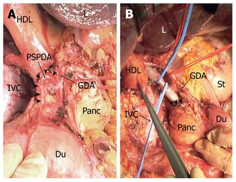
Figure 3 Photographs taken during an “extended” portal lymph node dissection.
A: The posterior superior pancreaticoduodenal artery (PSPDA; arrowheads) is shown following dissection of the posterior superior pancreaticoduodenal nodes. The common hepatic artery is secured with the red loop; B: The superior border of the uncinate process (arrowheads) is exposed, ensuring that dissection of the retroportal nodes is complete. The common bile duct was already transected at the level of PSPDA. The blue loops secure the portal vein, whereas the red loops secure the hepatic arteries. A forceps is picking up the node-bearing adipose tissues dissected en bloc. L: Liver; HDL: Hepatoduodenal ligament; GDA: Gastroduodenal artery; IVC: Inferior vena cava; Panc: Head of the pancreas; Du: Duodenum; St: Stomach.
- Citation: Shirai Y, Wakai T, Sakata J, Hatakeyama K. Regional lymphadenectomy for gallbladder cancer: Rational extent, technical details, and patient outcomes. World J Gastroenterol 2012; 18(22): 2775-2783
- URL: https://www.wjgnet.com/1007-9327/full/v18/i22/2775.htm
- DOI: https://dx.doi.org/10.3748/wjg.v18.i22.2775


