Copyright
©2012 Baishideng Publishing Group Co.
World J Gastroenterol. Jun 14, 2012; 18(22): 2775-2783
Published online Jun 14, 2012. doi: 10.3748/wjg.v18.i22.2775
Published online Jun 14, 2012. doi: 10.3748/wjg.v18.i22.2775
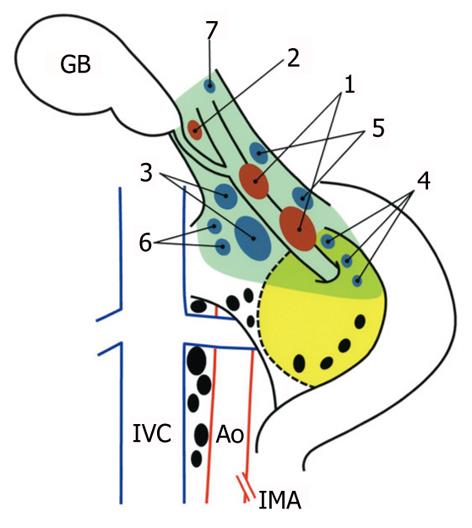
Figure 1 Topographical distribution of the regional lymph nodes of the gallbladder, shown after a full Kocher maneuver.
Solid ellipses represent individual lymph nodes: first-echelon nodes (red), second-echelon nodes (blue), and more distant nodes (black). The yellow painted area represents the head of the pancreas, and the dashed line indicates the border of the uncinate process of the pancreas. Arabic numerals indicate each of the first- and second-echelon node groups: (1) pericholedochal, nodes along the common bile duct; (2) cystic duct, node(s) along the cystic duct; (3) retroportal, nodes posterior to the portal vein and cephalad to the uncinate process; (4) posterior superior pancreaticoduodenal, nodes on the posterosuperior aspect of the head of the pancreas; (5) hepatic artery, nodes along the common or proper hepatic artery; (6) right celiac, nodes located right of the celiac axis and posterior to the common hepatic artery; and (7) hilar, nodes within the porta hepatis. The pale green area indicates the extent of a typical “extended” portal lymph node dissection (en bloc removal of the first- and second-echelon node groups) in the study department. GB: Gallbladder; IVC: Inferior vena cava; Ao: Aorta; IMA: Inferior mesenteric artery.
- Citation: Shirai Y, Wakai T, Sakata J, Hatakeyama K. Regional lymphadenectomy for gallbladder cancer: Rational extent, technical details, and patient outcomes. World J Gastroenterol 2012; 18(22): 2775-2783
- URL: https://www.wjgnet.com/1007-9327/full/v18/i22/2775.htm
- DOI: https://dx.doi.org/10.3748/wjg.v18.i22.2775


