Copyright
©2012 Baishideng Publishing Group Co.
World J Gastroenterol. May 7, 2012; 18(17): 2140-2144
Published online May 7, 2012. doi: 10.3748/wjg.v18.i17.2140
Published online May 7, 2012. doi: 10.3748/wjg.v18.i17.2140
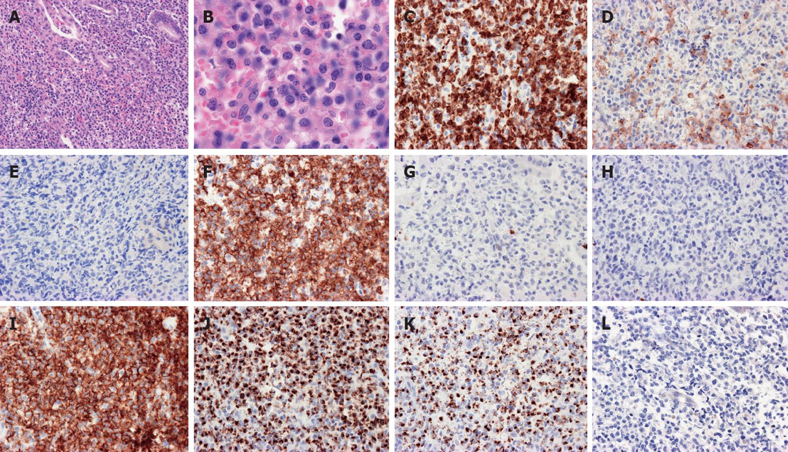
Figure 2 Histological examination showed massive atypical medium- to large-sized natural killer lymphocyte infiltrations with slightly irregular nuclear contours.
A dispersed chromatin pattern and clear cytoplasm in the gastric mucosa, × 100 (A) and × 400 (B); Immunohistochemical stains showed CD3+ (C), CD4- (D), CD5- (E), CD7+ (F), CD8- (G), CD20- (H), CD56+ (I), T-cell restricted intracellular antigen-1+ (J), granzyme B+ (K) and Epstein-Barr virus-encoded RNA in-situ hybridization (L).
- Citation: Terai T, Sugimoto M, Uozaki H, Kitagawa T, Kinoshita M, Baba S, Yamada T, Osawa S, Sugimoto K. Lymphomatoidgastropathy mimicking extranodal NK/T cell lymphoma, nasal type: A case report. World J Gastroenterol 2012; 18(17): 2140-2144
- URL: https://www.wjgnet.com/1007-9327/full/v18/i17/2140.htm
- DOI: https://dx.doi.org/10.3748/wjg.v18.i17.2140


