Copyright
©2012 Baishideng Publishing Group Co.
World J Gastroenterol. Apr 28, 2012; 18(16): 1968-1974
Published online Apr 28, 2012. doi: 10.3748/wjg.v18.i16.1968
Published online Apr 28, 2012. doi: 10.3748/wjg.v18.i16.1968
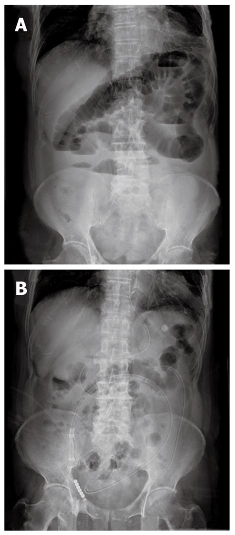
Figure 2 Radiographs of ileus tube decompression.
Plain abdominal radiographs (A) and (B) reviewed 3 d after ileus tube decompression compared with scans on admission in a patient with postoperative adhesive small bowel obstruction. A: The diffuse distended loops of small bowel that was filled with gas and fluid before intubation; air-fluid levels were seen in the enteric cavity; B: Reviewed 3 d after intubation; the previous gas-filled or fluid-filled small bowel loops showed no evidence of distention, the air-fluid levels disappeared, and the long tube had moved downward while the tip reached the distal jejunum.
-
Citation: Chen XL, Ji F, Lin Q, Chen YP, Lin JJ, Ye F, Yu JR, Wu YJ. A prospective randomized trial of transnasal ileus tube
vs nasogastric tube for adhesive small bowel obstruction. World J Gastroenterol 2012; 18(16): 1968-1974 - URL: https://www.wjgnet.com/1007-9327/full/v18/i16/1968.htm
- DOI: https://dx.doi.org/10.3748/wjg.v18.i16.1968


