Copyright
©2012 Baishideng Publishing Group Co.
World J Gastroenterol. Apr 7, 2012; 18(13): 1485-1495
Published online Apr 7, 2012. doi: 10.3748/wjg.v18.i13.1485
Published online Apr 7, 2012. doi: 10.3748/wjg.v18.i13.1485
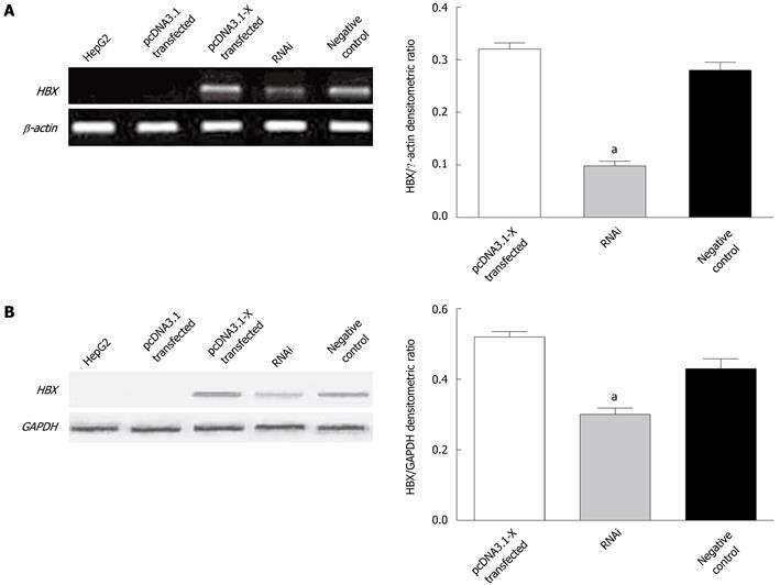
Figure 1 Detection of hepatitis B virus X protein expression in transfected HepG2 cells.
HepG2 cells were transfected with pcDNA3.1-X plasmids or cotransfected with pcDNA3.1-X and pSilencer3.1-shHBX plasmids. Forty-eight hours later, the expression of hepatitis B virus X protein (HBx) in HepG2 cells was determined by reverse transcription polymerase chain reaction (A) and Western blotting analysis (B). HepG2 group was not transfected with any plasmids. pcDNA3.1 transfected group was transfected with plasmid pcDNA3.1; pcDNA3.1-X transfected group was transfected with pcDNA3.1-X; RNAi group was cotransfected with pcDNA3.1-X and pSilencer3.1-shHBX in a ratio of 1:3; negative control group was cotransfected with pcDNA3.1-X and negative control plasmid pSilencer3.1-H1 in a ratio of 1:3. Data are expressed as mean ± SD (n = 3), aP < 0.05 vs pcDNA3.1-X transfected group.
- Citation: Tang RX, Kong FY, Fan BF, Liu XM, You HJ, Zhang P, Zheng KY. HBx activates FasL and mediates HepG2 cell apoptosis through MLK3-MKK7-JNKs signal module. World J Gastroenterol 2012; 18(13): 1485-1495
- URL: https://www.wjgnet.com/1007-9327/full/v18/i13/1485.htm
- DOI: https://dx.doi.org/10.3748/wjg.v18.i13.1485


