Copyright
©2012 Baishideng Publishing Group Co.
World J Gastroenterol. Apr 7, 2012; 18(13): 1438-1447
Published online Apr 7, 2012. doi: 10.3748/wjg.v18.i13.1438
Published online Apr 7, 2012. doi: 10.3748/wjg.v18.i13.1438
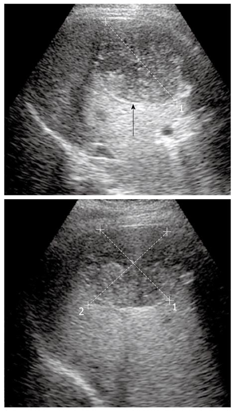
Figure 3 Ultrasonography images of hydatid disease show multiple internal septa and floating membranes inside the cyst.
Note the cyst wall is visible as double echogenic lines (see the black arrow).
- Citation: Marrone G, Crino' F, Caruso S, Mamone G, Carollo V, Milazzo M, Gruttadauria S, Luca A, Gridelli B. Multidisciplinary imaging of liver hydatidosis. World J Gastroenterol 2012; 18(13): 1438-1447
- URL: https://www.wjgnet.com/1007-9327/full/v18/i13/1438.htm
- DOI: https://dx.doi.org/10.3748/wjg.v18.i13.1438


