Copyright
©2011 Baishideng Publishing Group Co.
World J Gastroenterol. Mar 7, 2011; 17(9): 1192-1198
Published online Mar 7, 2011. doi: 10.3748/wjg.v17.i9.1192
Published online Mar 7, 2011. doi: 10.3748/wjg.v17.i9.1192
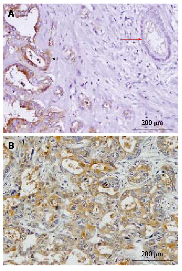
Figure 1 Representative immunohistochemical staining for CD133 in cholangiocarcinoma specimens.
A: Normal bile duct cells (red arrow) demonstrated negative staining, whereas cholangiocarcinoma cells had strong cytoplasmic staining (black arrow); B: Moderately differentiated cholangiocarcinoma with overexpression of CD133. Positive staining was observed in the cytoplasm of cholangiocarcinoma cells (200 × magnification).
- Citation: Leelawat K, Thongtawee T, Narong S, Subwongcharoen S, Treepongkaruna SA. Strong expression of CD133 is associated with increased cholangiocarcinoma progression. World J Gastroenterol 2011; 17(9): 1192-1198
- URL: https://www.wjgnet.com/1007-9327/full/v17/i9/1192.htm
- DOI: https://dx.doi.org/10.3748/wjg.v17.i9.1192


