Copyright
©2011 Baishideng Publishing Group Co.
World J Gastroenterol. Dec 28, 2011; 17(48): 5267-5273
Published online Dec 28, 2011. doi: 10.3748/wjg.v17.i48.5267
Published online Dec 28, 2011. doi: 10.3748/wjg.v17.i48.5267
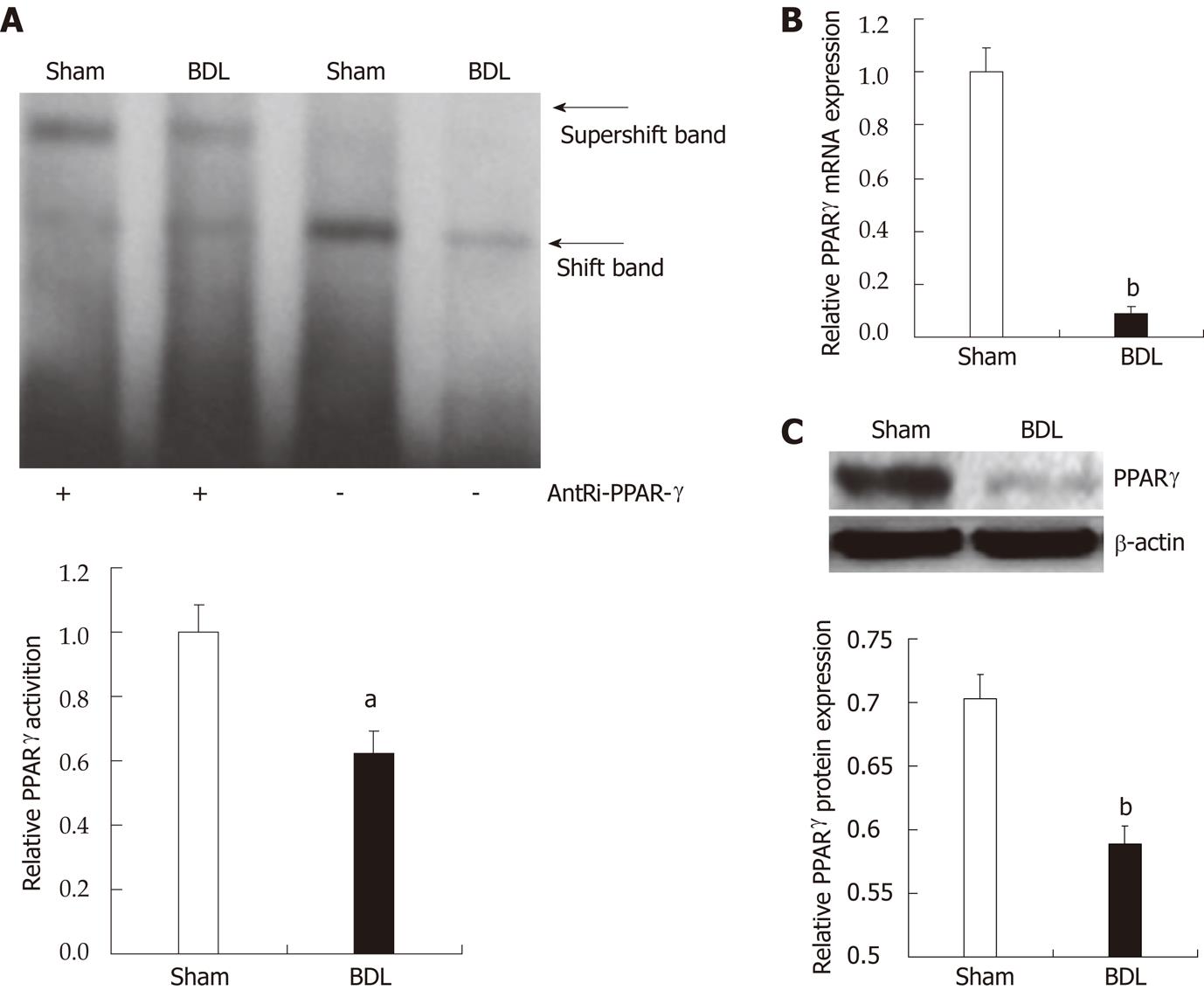
Figure 1 Modified measurement of peroxisome proliferator-activated receptor-γ activation and expression upon bile duct ligation and consequent cholestasis.
A: Electrophoretic mobility shift assay (top panel) of peroxisome proliferator-activated receptor (PPAR)-γ. DNA binding in nuclear extracts from liver, and relative densitometric analysis (lower panel). Data are expressed as mean ± SE (n = 3). aP < 0.05 for bile duct ligation (BDL) group vs sham group; B: PPAR-γ mRNA expression in the liver. PPAR-γ expression in the liver of BDL rats decreased significantly compared with the sham-group determined by quantitative real-time reverse transcription-polymerase chain reaction. Data are expressed as mean ± SE (n = 6). bP < 0.01 for BDL group vs sham group; C: Western blotting analysis (top panel) of PPAR-γ in nuclear extracts from liver, and relative densitometric analysis (lower panel, n = 3). Data are expressed as mean ± SE (n = 3). bP < 0.01 for BDL group vs sham group.
- Citation: Lv X, Song JG, Li HH, Ao JP, Zhang P, Li YS, Song SL, Wang XR. Decreased hepatic peroxisome proliferator-activated receptor-γ contributes to increased sensitivity to endotoxin in obstructive jaundice. World J Gastroenterol 2011; 17(48): 5267-5273
- URL: https://www.wjgnet.com/1007-9327/full/v17/i48/5267.htm
- DOI: https://dx.doi.org/10.3748/wjg.v17.i48.5267


