Copyright
©2011 Baishideng Publishing Group Co.
World J Gastroenterol. Nov 21, 2011; 17(43): 4757-4771
Published online Nov 21, 2011. doi: 10.3748/wjg.v17.i43.4757
Published online Nov 21, 2011. doi: 10.3748/wjg.v17.i43.4757
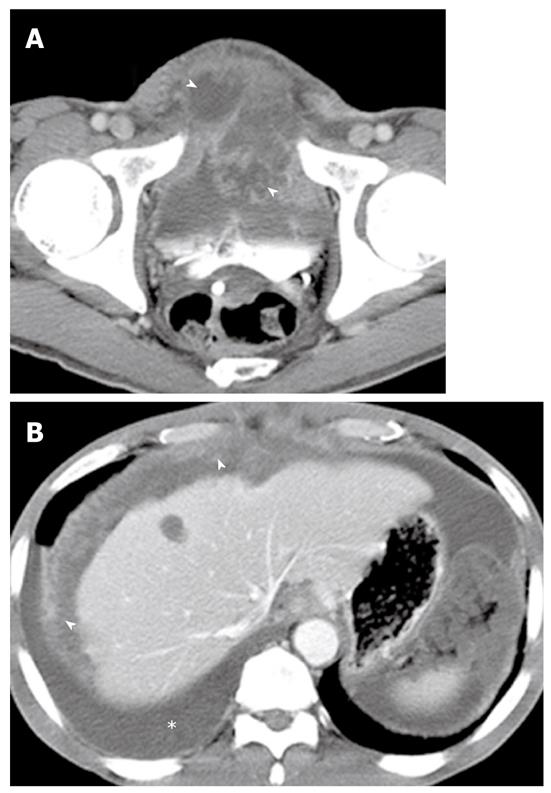
Figure 17 Intraperitoneal spread of mucinous adenocarcinoma of the urachus (also called peritoneal mucinous carcinomatosis) in a 57-year-old man.
A: Contrast-enhanced CT image shows a midline supravesical mass with heterogeneous attenuation. Within the mass are scattered low-attenuation areas (arrowheads), which represent mucin; B: Contrast-enhanced CT image of the upper abdomen shows low-attenuation mucinous ascites scalloping the liver margin. Note the right pleural effusion (asterisk) and diffuse nodular thickening of the peritoneum (arrowheads). CT: Computed tomography; MR: Magnetic resonance.
- Citation: Lee NK, Kim S, Kim HS, Jeon TY, Kim GH, Kim DU, Park DY, Kim TU, Kang DH. Spectrum of mucin-producing neoplastic conditions of the abdomen and pelvis: Cross-sectional imaging evaluation. World J Gastroenterol 2011; 17(43): 4757-4771
- URL: https://www.wjgnet.com/1007-9327/full/v17/i43/4757.htm
- DOI: https://dx.doi.org/10.3748/wjg.v17.i43.4757


