Copyright
©2011 Baishideng Publishing Group Co.
World J Gastroenterol. Nov 21, 2011; 17(43): 4757-4771
Published online Nov 21, 2011. doi: 10.3748/wjg.v17.i43.4757
Published online Nov 21, 2011. doi: 10.3748/wjg.v17.i43.4757
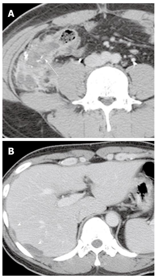
Figure 8 Mucinous cystadenocarcinoma of the appendix in a 26-year-old man.
A: Contrast-enhanced CT image shows a complex hypoattenuating mass with enhancing solid portions and punctate calcifications in the appendix (arrow); B: Contrast-enhanced CT image of the upper abdomen shows loculations of fluid scalloping on the liver surface (arrowheads), providing evidence of a mass effect. CT: Computed tomography.
- Citation: Lee NK, Kim S, Kim HS, Jeon TY, Kim GH, Kim DU, Park DY, Kim TU, Kang DH. Spectrum of mucin-producing neoplastic conditions of the abdomen and pelvis: Cross-sectional imaging evaluation. World J Gastroenterol 2011; 17(43): 4757-4771
- URL: https://www.wjgnet.com/1007-9327/full/v17/i43/4757.htm
- DOI: https://dx.doi.org/10.3748/wjg.v17.i43.4757


