Copyright
©2011 Baishideng Publishing Group Co.
World J Gastroenterol. Oct 14, 2011; 17(38): 4334-4338
Published online Oct 14, 2011. doi: 10.3748/wjg.v17.i38.4334
Published online Oct 14, 2011. doi: 10.3748/wjg.v17.i38.4334
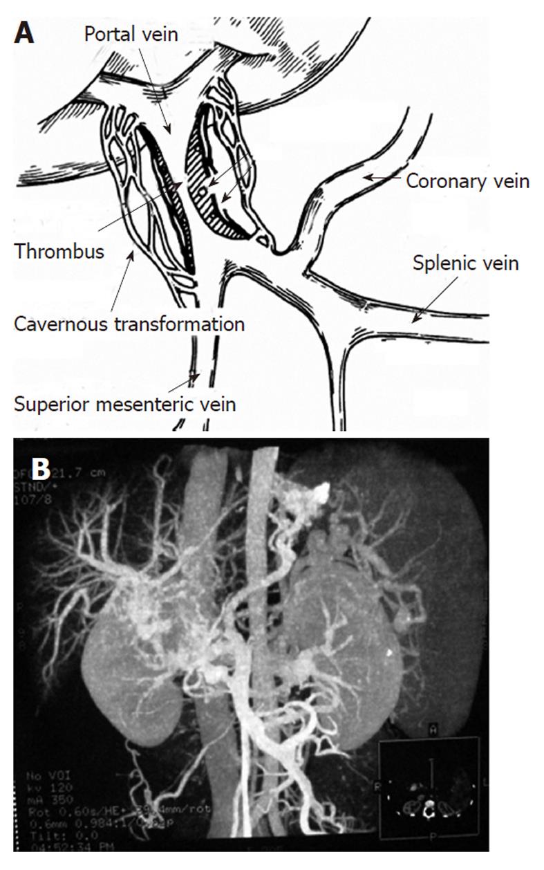
Figure 1 Cavernous transformation of portal vein and sixty-four-slice computed tomography angiography.
A: Illustration of cavernous transformation of the portal vein (PV); B: Sixty-four-slice computed tomography angiography of cavernous transformation in the extrahepatic PV indicates varices in the esophagus and gastrosplenic area.
- Citation: Zhang MM, Pu CL, Li YC, Guo CB. Sixty-four-slice computed tomography in surgical strategy of portal vein cavernous transformation. World J Gastroenterol 2011; 17(38): 4334-4338
- URL: https://www.wjgnet.com/1007-9327/full/v17/i38/4334.htm
- DOI: https://dx.doi.org/10.3748/wjg.v17.i38.4334


