Copyright
©2011 Baishideng Publishing Group Co.
World J Gastroenterol. Sep 28, 2011; 17(36): 4076-4089
Published online Sep 28, 2011. doi: 10.3748/wjg.v17.i36.4076
Published online Sep 28, 2011. doi: 10.3748/wjg.v17.i36.4076
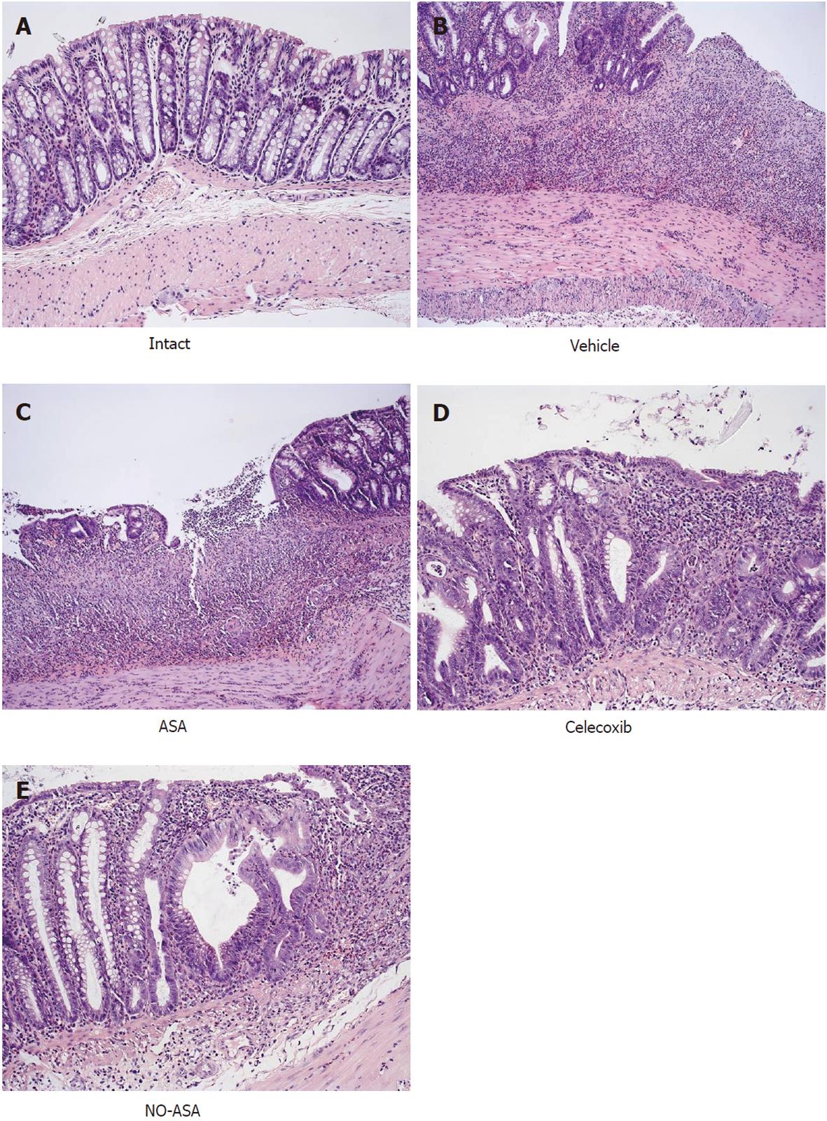
Figure 5 Histological appearance of the intact colonic mucosa (A) and that treated with vehicle (B), aspirin (C), celecoxib (D) and nitric oxide releasing aspirin on day 10 in rats with trinitrobenzenesulfonic acid-induced colitis.
Intact rat colon shows regular colonic architecture and normal colonic mucosa continuity with no signs of inflammation (A). In trinitrobenzenesulfonic acid rats treated with vehicle, deep ulceration and an intense neutrophil infiltration with the presence of numerous neutrophils penetrating the muscularis mucosa and submucosa were observed. The features of regeneration adjacent to the ulcer margin are clearly visible. In aspirin (ASA)-treated rats with colitis (C), there is deep ulceration with necrosis and an intense inflammation followed by severe neutrophil infiltration and the formation of granulation tissue penetrating the muscle layer of the muscularis propria. Less regeneration is observed with ASA (C) than vehicle (control) animals (B). The partially healed epithelium and abnormal crypt architecture with much more pronounced regeneration was observed in the colonic mucosa of rats with colitis treated with celecoxib (D) as compared with other cyclooxygenase inhibitors. In nitric oxide (NO)-ASA treated rats with colitis (E), the most advanced healing process of colonic ulcers as reflected by scar formation, epithelial regeneration and significantly smaller neutrophil infiltration was observed. Part of the colonic crypts distant to the scar already shows a normal appearance.
- Citation: Zwolinska-Wcislo M, Brzozowski T, Ptak-Belowska A, Targosz A, Urbanczyk K, Kwiecien S, Sliwowski Z. Nitric oxide-releasing aspirin but not conventional aspirin improves healing of experimental colitis. World J Gastroenterol 2011; 17(36): 4076-4089
- URL: https://www.wjgnet.com/1007-9327/full/v17/i36/4076.htm
- DOI: https://dx.doi.org/10.3748/wjg.v17.i36.4076


