Copyright
©2011 Baishideng Publishing Group Co.
World J Gastroenterol. Sep 14, 2011; 17(34): 3922-3932
Published online Sep 14, 2011. doi: 10.3748/wjg.v17.i34.3922
Published online Sep 14, 2011. doi: 10.3748/wjg.v17.i34.3922
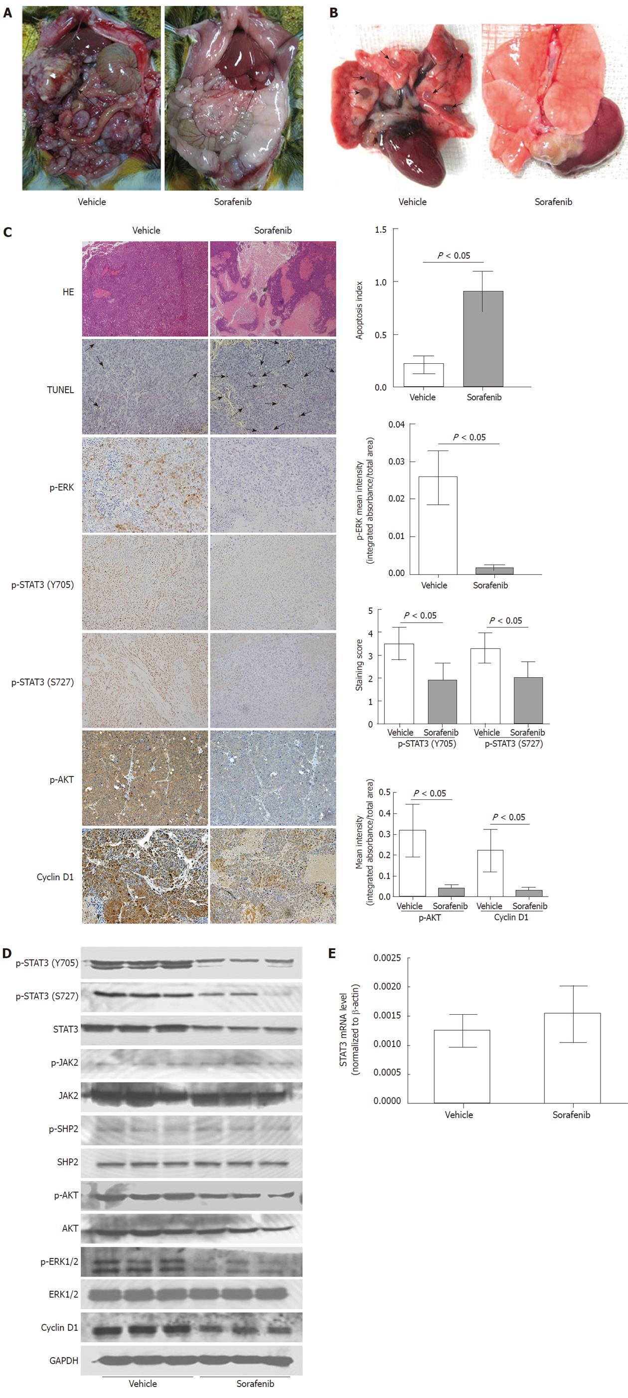
Figure 3 Inhibition of tumor growth and metastasis by sorafenib in vivo.
Rats with morris hepatoma (MH) tumors were treated with vehicle or sorafenib (30 mg/kg per day) starting on day 17 after tumor implantation. Tumors were removed on day 38 following implantation; A: Tumor growth and abdominal lymph node metastasis were reduced by sorafenib treatment (arrows); B: Sorafenib treatment reduced lung metastasis (arrows); C: Significant tumor necrosis in the sorafenib-treated group was visualized by hematoxylin-eosin staining (magnification, × 40). As shown by TUNEL (arrows), sorafenib also induced tumor cell apoptosis significantly (apoptosis index, 0.217 ± 0.825 vs 0.909 ± 0.189; P < 0.05; magnification, × 200). Immunohistochemical analysis showed that the expression levels of cyclin D1 and phosphorylation of signal transducer and activator of transcription 3 (STAT3) (Y705 and S727), Akt, and extracellular signal-regulated kinase (ERK) were significantly reduced by sorafenib treatment (P < 0.05 for all; magnification, × 200); D: Sorafenib reduced phosphorylation of STAT3, Akt, and ERK, but not Janus kinase 2 and shatterproof 2. Sorafenib also reduced the expression levels of cyclin D1 significantly; E: Sorafenib did not affect STAT3 mRNA levels in rat tumor tissues (P > 0.05).
- Citation: Gu FM, Li QL, Gao Q, Jiang JH, Huang XY, Pan JF, Fan J, Zhou J. Sorafenib inhibits growth and metastasis of hepatocellular carcinoma by blocking STAT3. World J Gastroenterol 2011; 17(34): 3922-3932
- URL: https://www.wjgnet.com/1007-9327/full/v17/i34/3922.htm
- DOI: https://dx.doi.org/10.3748/wjg.v17.i34.3922


