Copyright
©2011 Baishideng Publishing Group Co.
World J Gastroenterol. Aug 21, 2011; 17(31): 3614-3622
Published online Aug 21, 2011. doi: 10.3748/wjg.v17.i31.3614
Published online Aug 21, 2011. doi: 10.3748/wjg.v17.i31.3614
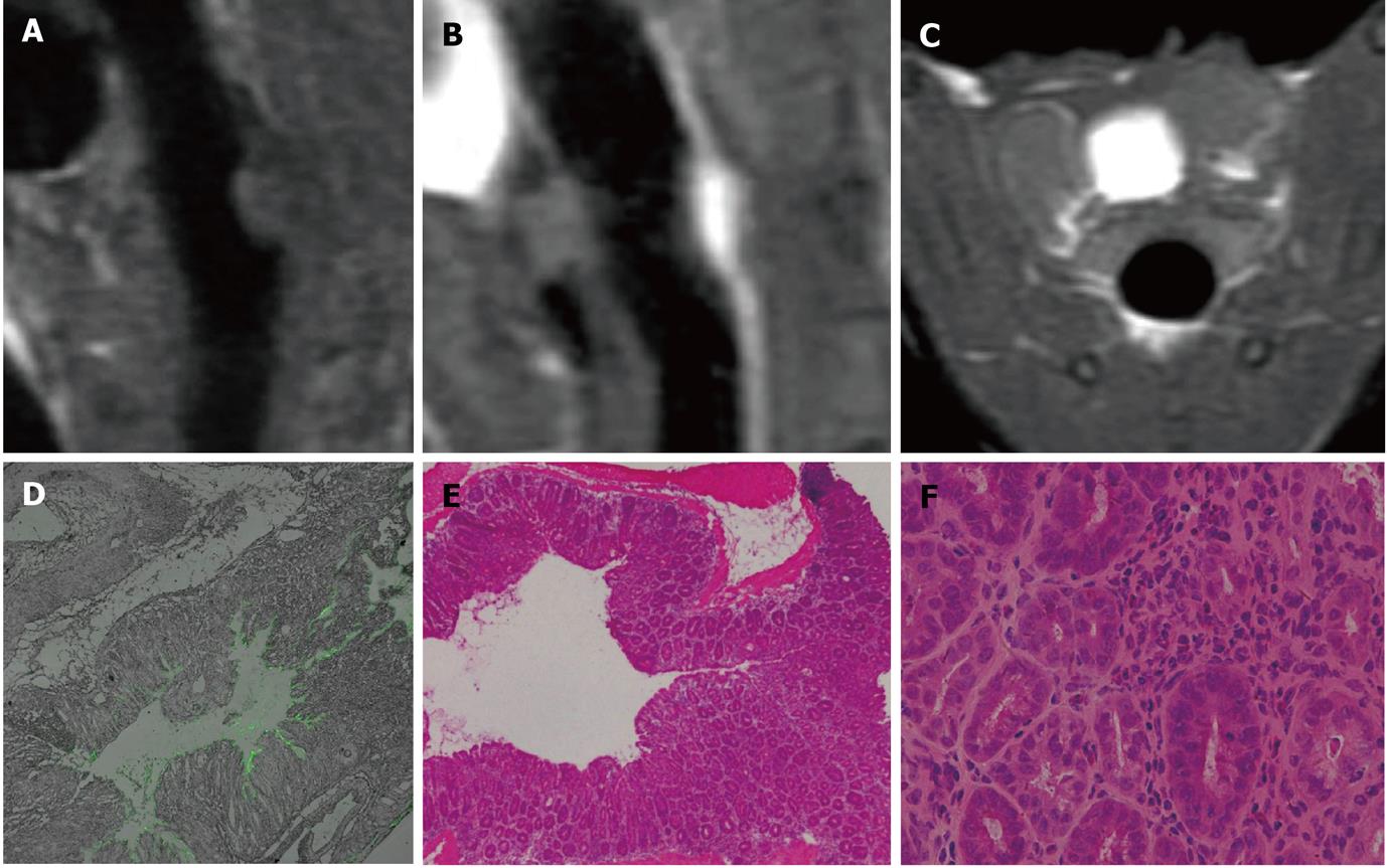
Figure 6 Peri-tumor interstitial fluorescein isothiocyanate deposition.
A: A broad-based mass (high-level adenoma) in the pre-enema fluid attenuated inversion recovery (FLAIR) image; B, C: Tumor and peri-tumor mural enhancement, together with bladder imaging agent accumulation, in the post-enema FLAIR image; D: Interstitial linear fluorescein isothiocyanate deposition in the peri-tumor colorectal wall; E, F: Hematoxylin and eosin images at the same slice, obvious nuclear atypia identified.
- Citation: Wu T, Zheng WL, Zhang SZ, Sun JH, Yuan H. Bimodal visualization of colorectal uptake of nanoparticles in dimethylhydrazine-treated mice. World J Gastroenterol 2011; 17(31): 3614-3622
- URL: https://www.wjgnet.com/1007-9327/full/v17/i31/3614.htm
- DOI: https://dx.doi.org/10.3748/wjg.v17.i31.3614


