Copyright
©2011 Baishideng Publishing Group Co.
World J Gastroenterol. Aug 14, 2011; 17(30): 3544-3553
Published online Aug 14, 2011. doi: 10.3748/wjg.v17.i30.3544
Published online Aug 14, 2011. doi: 10.3748/wjg.v17.i30.3544
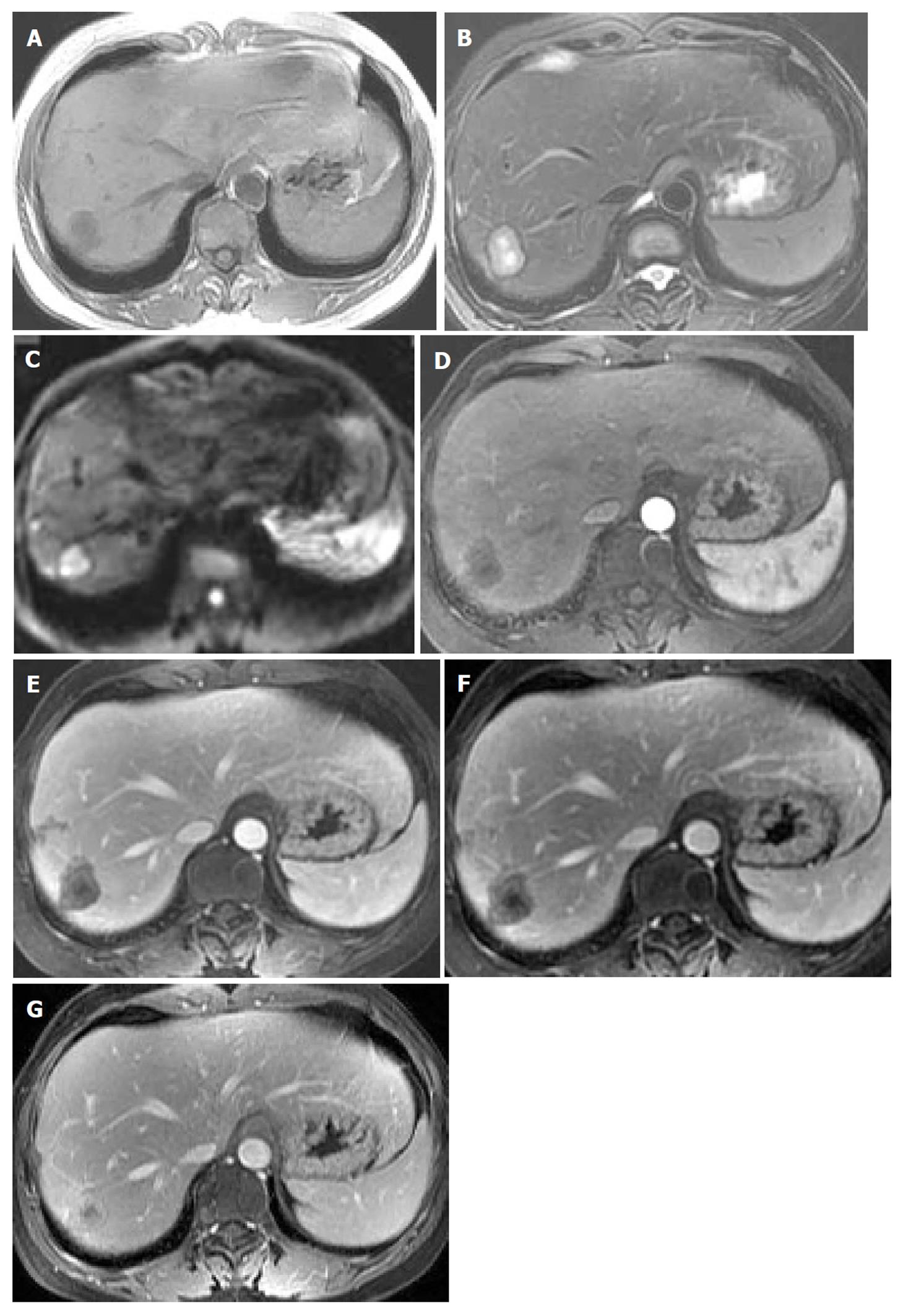
Figure 2 Multifocal hepatic epithelioid hemangioendothelioma in a 48-year-old female.
Precontrast axial magnetic resonance imaging scan of the liver shows multiple lesions of low signal intensity with peripheral faint hyperintensity on T1WI (A), high signal intensity with peripheral hypointensity and an area of evident hyperintensity in the center of one lesion on T2WI (B), and hyperintensity with peripheral hypointensity on diffusion weighted imaging (C). Lesions show peripheral ring-like enhancement in the arterial phase (D), and heterogeneously progressive reinforcement in the portal venous phase (E), equilibrium phase (F) and it approaches isointensity to liver parenchyma in the delayed phase (G). There is an association with an area of unenhanced necrosis in the center, which looks like a conspicuous “target” with an inner low intensity, in-between high intensity and outer lower intensity layers.
- Citation: Chen Y, Yu RS, Qiu LL, Jiang DY, Tan YB, Fu YB. Contrast-enhanced multiple-phase imaging features in hepatic epithelioid hemangioendothelioma. World J Gastroenterol 2011; 17(30): 3544-3553
- URL: https://www.wjgnet.com/1007-9327/full/v17/i30/3544.htm
- DOI: https://dx.doi.org/10.3748/wjg.v17.i30.3544


