
















©2011 Baishideng Publishing Group Co.
World J Gastroenterol. Jan 21, 2011; 17(3): 403-406
Published online Jan 21, 2011. doi: 10.3748/wjg.v17.i3.403
Published online Jan 21, 2011. doi: 10.3748/wjg.v17.i3.403
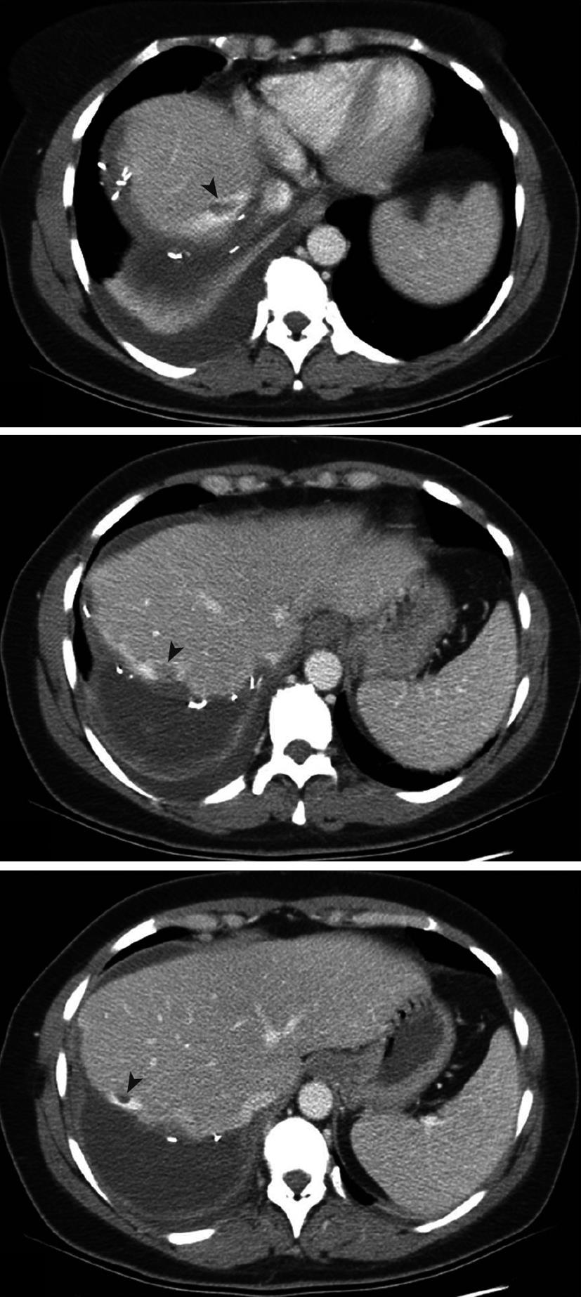
Figure 1 Postoperative computed tomography scan (day 4) in case 1.
Defects are present in the middle hepatic vein close to the transection plan (arrowheads).
- Citation: Buc E, Dokmak S, Zappa M, Denninger MH, Valla DC, Belghiti J, Farges O. Hepatic veins as a site of clot formation following liver resection. World J Gastroenterol 2011; 17(3): 403-406
- URL: https://www.wjgnet.com/1007-9327/full/v17/i3/403.htm
- DOI: https://dx.doi.org/10.3748/wjg.v17.i3.403