Copyright
©2011 Baishideng Publishing Group Co.
World J Gastroenterol. Jul 28, 2011; 17(28): 3322-3329
Published online Jul 28, 2011. doi: 10.3748/wjg.v17.i28.3322
Published online Jul 28, 2011. doi: 10.3748/wjg.v17.i28.3322
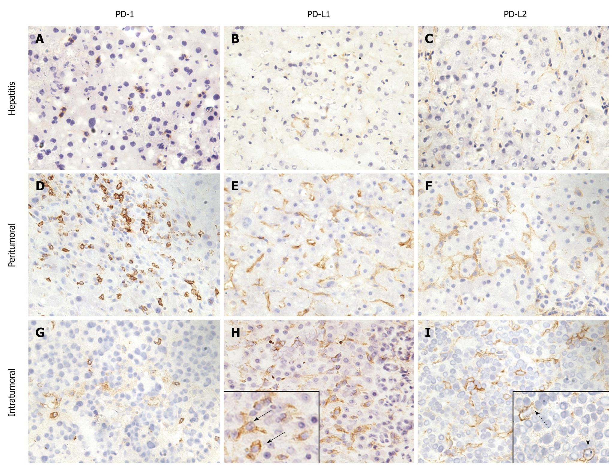
Figure 1 Immunohistochemical staining of liver tissues from patients with hepatitis and hepatocellular carcinoma for programmed death-1, programmed death-L1, and programmed death-L2.
A-C: Programmed death (PD)-1 (A), PD ligand 1 (PD-L1) (B), and PD-L2 (C) expression in liver tissues with hepatitis; D-I: PD-1 (D and G), PD-L1 (E and H), and PD-L2 (F and I) expression in liver tissues with hepatocellular carcinoma (HCC); D-F: Peritumoral region; G-I: Intratumoral region. A, D and G: PD-1 was expressed on the membrane of infiltrated lymphocytes in liver tissues with hepatitis and HCC; B, E and H: PD-L1 was expressed on the membrane of hepatic cells and/or tumor cells in liver tissues with hepatitis and HCC; C, F and I: PD-L2 was expressed on the membrane of hepatic cells and/or tumor cells in liver tissues with hepatitis and HCC. Solid arrows indicate PD-L1+ tumor cells, and dashed arrows indicate PD-L2+ tumor cells. Magnification 200 ×.
- Citation: Wang BJ, Bao JJ, Wang JZ, Wang Y, Jiang M, Xing MY, Zhang WG, Qi JY, Roggendorf M, Lu MJ, Yang DL. Immunostaining of PD-1/PD-Ls in liver tissues of patients with hepatitis and hepatocellular carcinoma. World J Gastroenterol 2011; 17(28): 3322-3329
- URL: https://www.wjgnet.com/1007-9327/full/v17/i28/3322.htm
- DOI: https://dx.doi.org/10.3748/wjg.v17.i28.3322


