Copyright
©2011 Baishideng Publishing Group Co.
World J Gastroenterol. Jul 7, 2011; 17(25): 3060-3065
Published online Jul 7, 2011. doi: 10.3748/wjg.v17.i25.3060
Published online Jul 7, 2011. doi: 10.3748/wjg.v17.i25.3060
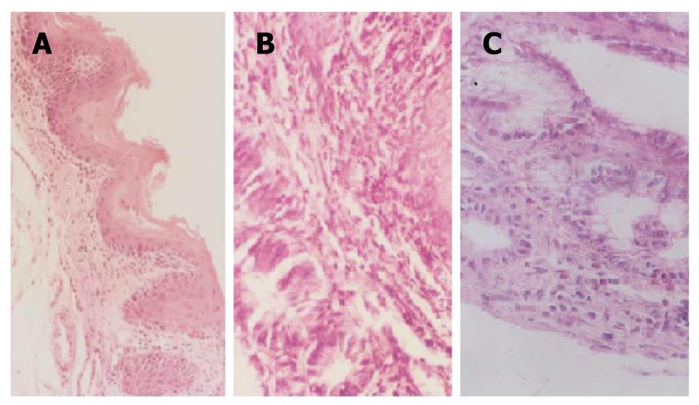
Figure 4 The changes of the esophagus mucosa in the sham operated group and the animal model groups under the light microscope (200 ×).
A: Normal esophagus in the sham operated group; B: Esophagitis and barrett esophagus in the model group; C: Intestinal metaplasia with dysplasia and esophageal adenocarcinoma in the model group.
- Citation: Cheng P, Li JS, Gong J, Zhang LF, Chen RZ. Effects of refluxate pH values on duodenogastroesophageal reflux-induced esophageal adenocarcinoma. World J Gastroenterol 2011; 17(25): 3060-3065
- URL: https://www.wjgnet.com/1007-9327/full/v17/i25/3060.htm
- DOI: https://dx.doi.org/10.3748/wjg.v17.i25.3060


