Copyright
©2011 Baishideng Publishing Group Co.
World J Gastroenterol. Jun 28, 2011; 17(24): 2924-2932
Published online Jun 28, 2011. doi: 10.3748/wjg.v17.i24.2924
Published online Jun 28, 2011. doi: 10.3748/wjg.v17.i24.2924
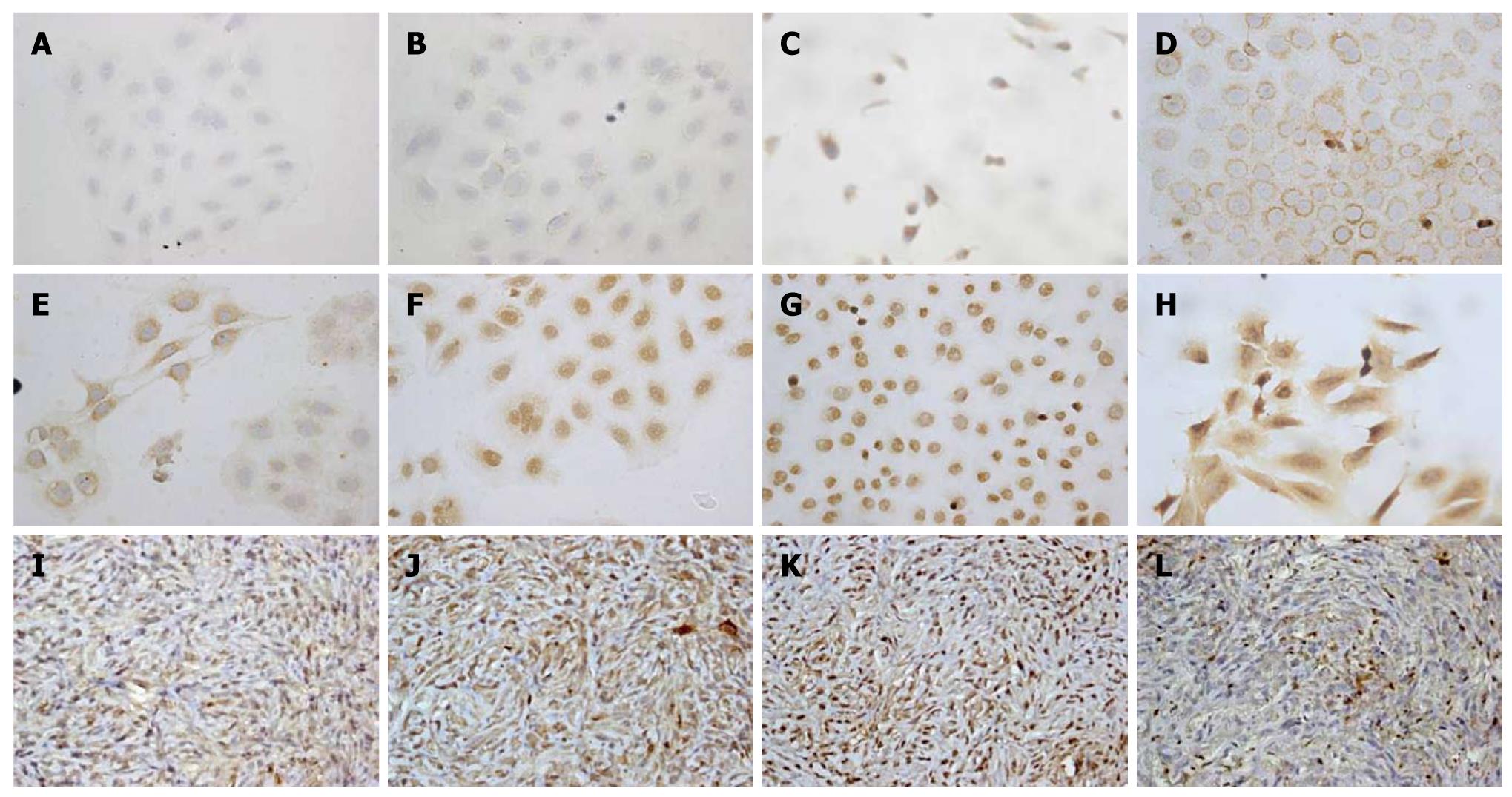
Figure 4 Immunohistochemical staining (× 400) analysis of Chang Gung CCA cells (upper two panels, A-H) and xenograft tissues of CCA mouse models (lower panel, I-L).
A: Negative control; B: Negative expression of K-ras; C: EGFR weakly expressed in a cytoplasmic distribution; D-G: Her-II, Biliary cytokeratin (CK19), COX-II, and Met diffusely expressed in a cytoplasmic distribution; H: MUC-4 strongly and diffusely expressed in a cytoplasmic and membranous distribution; I-L: The results revealed that CK-19, COX-II, Met, and MUC4 are diffusely expressed in a cytoplasmic distribution in the rat CCA xenograft.
- Citation: Yeh CN, Lin KJ, Chen TW, Wu RC, Tsao LC, Chen YT, Weng WH, Chen MF. Characterization of a novel rat cholangiocarcinoma cell culture model-CGCCA. World J Gastroenterol 2011; 17(24): 2924-2932
- URL: https://www.wjgnet.com/1007-9327/full/v17/i24/2924.htm
- DOI: https://dx.doi.org/10.3748/wjg.v17.i24.2924


