Copyright
©2011 Baishideng Publishing Group Co.
World J Gastroenterol. Jun 7, 2011; 17(21): 2667-2673
Published online Jun 7, 2011. doi: 10.3748/wjg.v17.i21.2667
Published online Jun 7, 2011. doi: 10.3748/wjg.v17.i21.2667
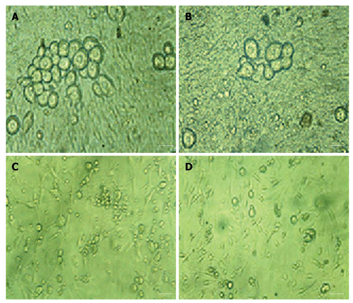
Figure 5 Morphological changes in dorsal root ganglion neurons.
A: Monoculture of dorsal root ganglion (DRG) neurons on the 1st d (× 400); B: Monoculture of DRG neurons on the 7th d (× 400); C: Co-culture of DRG neurons and infected BxPC-3 cells (× 200); D: Co-culture of DRG neurons and normal BxPC-3 cells (× 200). Scale bars: 25 μm (A, B) and 50 μm (C, D).
- Citation: Yao J, Zhang M, Ma QY, Wang Z, Wang LC, Zhang D. PAd-shRNA-PTN reduces pleiotrophin of pancreatic cancer cells and inhibits neurite outgrowth of DRG. World J Gastroenterol 2011; 17(21): 2667-2673
- URL: https://www.wjgnet.com/1007-9327/full/v17/i21/2667.htm
- DOI: https://dx.doi.org/10.3748/wjg.v17.i21.2667


