Copyright
©2011 Baishideng Publishing Group Co.
World J Gastroenterol. Jun 7, 2011; 17(21): 2585-2591
Published online Jun 7, 2011. doi: 10.3748/wjg.v17.i21.2585
Published online Jun 7, 2011. doi: 10.3748/wjg.v17.i21.2585
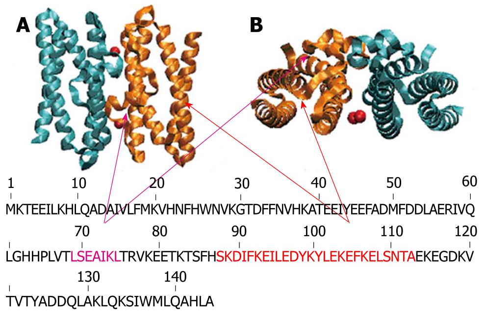
Figure 2 Schematic representation of exposed helices of Helicobacter pylori neutrophil activating protein.
Helicobacter pylori neutrophil activating protein dimer in stand up (A) and top view (B) with the exposed helices H3 and H4 (therefore possible candidates for interacting with the neutrophils) colored in violet and orange respectively.
-
Citation: Choli-Papadopoulou T, Kottakis F, Papadopoulos G, Pendas S.
Helicobacter pylori neutrophil activating protein as target for new drugs againstH. pylori inflammation. World J Gastroenterol 2011; 17(21): 2585-2591 - URL: https://www.wjgnet.com/1007-9327/full/v17/i21/2585.htm
- DOI: https://dx.doi.org/10.3748/wjg.v17.i21.2585


