Copyright
©2011 Baishideng Publishing Group Co.
World J Gastroenterol. Jan 14, 2011; 17(2): 213-218
Published online Jan 14, 2011. doi: 10.3748/wjg.v17.i2.213
Published online Jan 14, 2011. doi: 10.3748/wjg.v17.i2.213
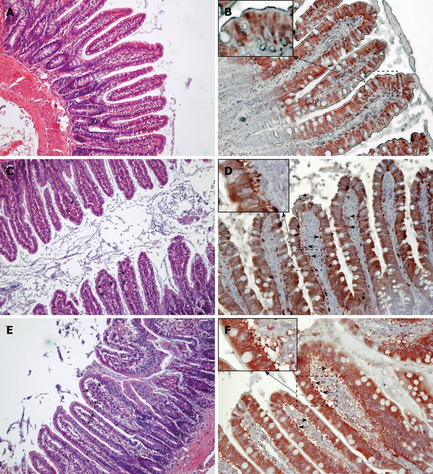
Figure 4 Histological and immunohistochemical evaluation of intestinal injury.
Histological evaluation of the ileum was performed using HE staining (A, C and E, × 100). When compared to the saline group (A, B), water (C, D) and free oxyhemoglobin (FHb) (E, F) infusions led to the development of subepithelial spaces (asterisks) in the villi. In support of the findings of plasmatic release of ileal lipid binding protein (iLBP), immunohistochemical analysis of the ileum shows cytosolic staining for iLBP in the epithelial cells of the upper part of the villi (B, × 200). Leaking of iLBP in the subepithelial spaces (arrows) indicates intestinal epithelial cell injury after water or FHb infusion (D, F, × 200). Insets show 400 × magnification of selected areas where staining of iLBP can be seen outside the intestinal epithelial cellular membrane, indicating epithelial cellular injury (D, F).
- Citation: Hanssen SJ, Lubbers T, Hodin CM, Prinzen FW, Buurman WA, Jacobs MJ. Hemolysis results in impaired intestinal microcirculation and intestinal epithelial cell injury. World J Gastroenterol 2011; 17(2): 213-218
- URL: https://www.wjgnet.com/1007-9327/full/v17/i2/213.htm
- DOI: https://dx.doi.org/10.3748/wjg.v17.i2.213


