Copyright
©2011 Baishideng Publishing Group Co.
World J Gastroenterol. Apr 21, 2011; 17(15): 1947-1960
Published online Apr 21, 2011. doi: 10.3748/wjg.v17.i15.1947
Published online Apr 21, 2011. doi: 10.3748/wjg.v17.i15.1947
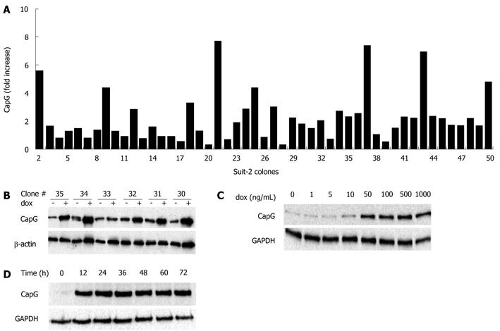
Figure 3 Selection (A, B) and inducibility (C, D) of stably-transfected Sβtet29Cap clones.
The stable Sβtet29 clone was transfected with the pTRE2hygCapG full size vector and clones selected with hygromycin B (200 μg/mL). A: Individual clones (n = 49) were induced with 500 ng/mL doxycycline for 24 h and the protein lysate subjected to Western blotting. The CapG level was calculated for each individual clone in the non-induced and the induced state. The fold increase (normalised to actin) in CapG for each of the 49 clones is shown in the bar chart; B: A representative Western blotting for CapG is shown for pTRE2hygCapG clones 30 to 35. The indicates the basal CapG expression, + doxycycline (dox) induced; C: The inducibility of CapG protein expression was investigated by treating the Sβtet29Cap35 cells with 0, 1, 5, 10, 50, 100, 500, 1000 ng/mL dox for 24 h. The resulting Western blotting for CapG and GAPDH as a loading control is shown; D: The Western blotting represents the CapG protein level of Sβtet29Cap35 cells, induced with 500 ng/mL doxycycline for 0, 12, 24, 36 and 48 h. GAPDH was used as a loading control.
- Citation: Tonack S, Patel S, Jalali M, Nedjadi T, Jenkins RE, Goldring C, Neoptolemos J, Costello E. Tetracycline-inducible protein expression in pancreatic cancer cells: Effects of CapG overexpression. World J Gastroenterol 2011; 17(15): 1947-1960
- URL: https://www.wjgnet.com/1007-9327/full/v17/i15/1947.htm
- DOI: https://dx.doi.org/10.3748/wjg.v17.i15.1947


