Copyright
©2011 Baishideng Publishing Group Co.
World J Gastroenterol. Mar 21, 2011; 17(11): 1427-1433
Published online Mar 21, 2011. doi: 10.3748/wjg.v17.i11.1427
Published online Mar 21, 2011. doi: 10.3748/wjg.v17.i11.1427
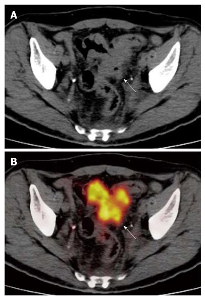
Figure 2 The computed tomography (A) and fused positron emission tomography/computed tomography (B) images show diffuse and irregular neoplastic thickening of sigmoid colon walls, which show rounded advancing margins with intense radiotracer uptake (arrows) (SUVmax 13).
The lesion was correctly classified as T3.
- Citation: Mainenti PP, Iodice D, Segreto S, Storto G, Magliulo M, Palma GDD, Salvatore M, Pace L. Colorectal cancer and 18FDG-PET/CT: What about adding the T to the N parameter in loco-regional staging? World J Gastroenterol 2011; 17(11): 1427-1433
- URL: https://www.wjgnet.com/1007-9327/full/v17/i11/1427.htm
- DOI: https://dx.doi.org/10.3748/wjg.v17.i11.1427


