Copyright
©2011 Baishideng Publishing Group Co.
World J Gastroenterol. Mar 14, 2011; 17(10): 1358-1361
Published online Mar 14, 2011. doi: 10.3748/wjg.v17.i10.1358
Published online Mar 14, 2011. doi: 10.3748/wjg.v17.i10.1358
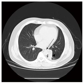
Figure 2 Preoperative chest computed tomography scan demonstrating abnormal communication between the lower third of esophagus and the left segmental bronchus (arrow) and a mass measuring 3.
0 cm in diameter with irregular borders encircling the basal segmental bronchi of the left lower lobe caused by bronchoesophageal fistula.
- Citation: Zhang BS, Zhou NK, Yu CH. Congenital bronchoesophageal fistula in adults. World J Gastroenterol 2011; 17(10): 1358-1361
- URL: https://www.wjgnet.com/1007-9327/full/v17/i10/1358.htm
- DOI: https://dx.doi.org/10.3748/wjg.v17.i10.1358


