Copyright
©2011 Baishideng Publishing Group Co.
World J Gastroenterol. Jan 7, 2011; 17(1): 53-62
Published online Jan 7, 2011. doi: 10.3748/wjg.v17.i1.53
Published online Jan 7, 2011. doi: 10.3748/wjg.v17.i1.53
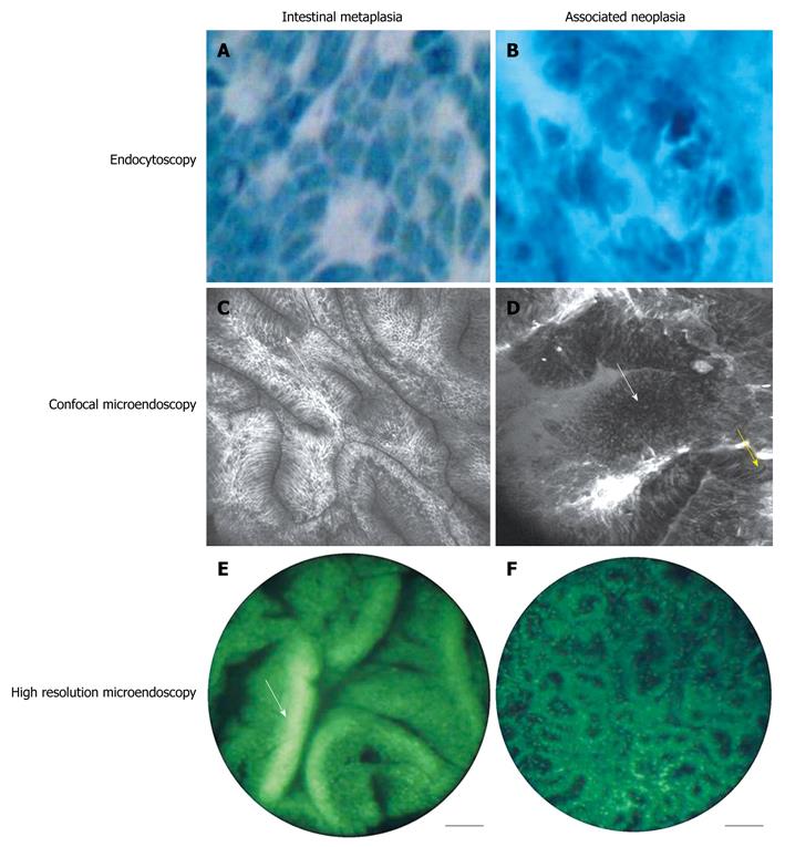
Figure 3 Images representing intestinal metaplasia and neoplasia collected using endocytoscopy (A, B) (Copyright (2007), with permission from Thieme)[47], confocal microendoscopy (C, D) [Copyright (2006), with permission from Elsevier][48], and high-resolution microendoscopy (E, F) [Copyright (2008), with permission from Elsevier][51].
Topically applied methylene blue is used in endocytoscopy to highlight nuclear changes (A, B); In metaplasia (A), nuclei appear organized and regular; this is in stark contrast to neoplasia (B) where nuclei appear pleomorphic. Both images were taken using 1125 × magnification. Confocal images were taken using intravenous fluorescein to enhance contrast of subepithelial capillaries (C, D); for intestinal metaplasia (C), confocal microendoscopy allows visualization of mucin-containing goblet cells (white arrow); For Barrett’s-associated neoplasia (B), cells are irregularly oriented (white arrow) and malignant invasion of the lamina propria can be seen (yellow arrow). Confocal images are 500 μm × 500 μm. High-resolution microendoscopy uses proflavine for contrast enhancement, highlighting changes in glandular and nuclear patterns (E, F). High-resolution images are 750 μm in diameter.
- Citation: Thekkek N, Anandasabapathy S, Richards-Kortum R. Optical molecular imaging for detection of Barrett’s-associated neoplasia. World J Gastroenterol 2011; 17(1): 53-62
- URL: https://www.wjgnet.com/1007-9327/full/v17/i1/53.htm
- DOI: https://dx.doi.org/10.3748/wjg.v17.i1.53


