Copyright
©2011 Baishideng Publishing Group Co.
World J Gastroenterol. Jan 7, 2011; 17(1): 28-41
Published online Jan 7, 2011. doi: 10.3748/wjg.v17.i1.28
Published online Jan 7, 2011. doi: 10.3748/wjg.v17.i1.28
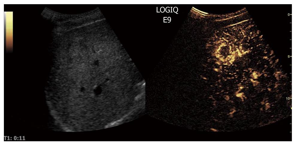
Figure 10 Contrast-enhanced ultrasound allows for visualization of the arteriogram of hepatocellular carcinoma in the early arterial phase.
The feeding vessel is visible on the tumor right side. Typically there is initial peripheral enhancement before the centripetal influx to the center of the tumor.
- Citation: Postema M, Gilja OH. Contrast-enhanced and targeted ultrasound. World J Gastroenterol 2011; 17(1): 28-41
- URL: https://www.wjgnet.com/1007-9327/full/v17/i1/28.htm
- DOI: https://dx.doi.org/10.3748/wjg.v17.i1.28


