Copyright
©2010 Baishideng.
World J Gastroenterol. Feb 28, 2010; 16(8): 1039-1042
Published online Feb 28, 2010. doi: 10.3748/wjg.v16.i8.1039
Published online Feb 28, 2010. doi: 10.3748/wjg.v16.i8.1039
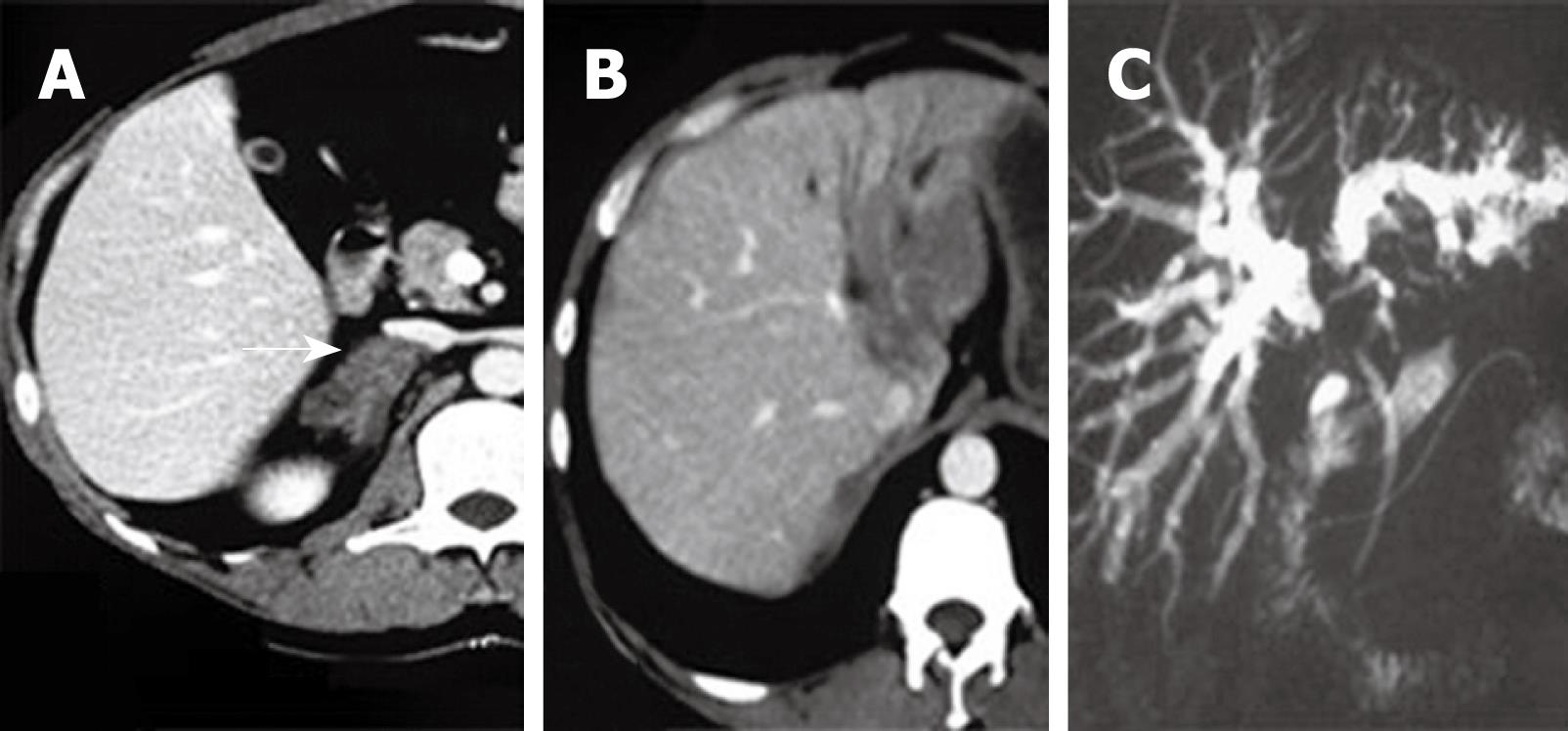
Figure 2 Imaging changes.
A: Arrow shows computed tomography (CT) features of adrenal mucormycosis; B: CT features of hepatic lesion. A well circumscribed hypodense lesion in hilar and left lateral lobe, surrounding the vessels without a mass effect, should suggest an angioinvasive organism. This lesion presents necrosis of liver tissue due to fungal thrombosis; C: Magnetic resonance cholangiopancreatography demonstrates an abrupt stenosis of the primary biliary confluence with symmetric upstream dilation of the intrahepatic bile ducts.
- Citation: Li KW, Wen TF, Li GD. Hepatic mucormycosis mimicking hilar cholangiocarcinoma: A case report and literature review. World J Gastroenterol 2010; 16(8): 1039-1042
- URL: https://www.wjgnet.com/1007-9327/full/v16/i8/1039.htm
- DOI: https://dx.doi.org/10.3748/wjg.v16.i8.1039


