Copyright
©2010 Baishideng.
World J Gastroenterol. Feb 14, 2010; 16(6): 683-690
Published online Feb 14, 2010. doi: 10.3748/wjg.v16.i6.683
Published online Feb 14, 2010. doi: 10.3748/wjg.v16.i6.683
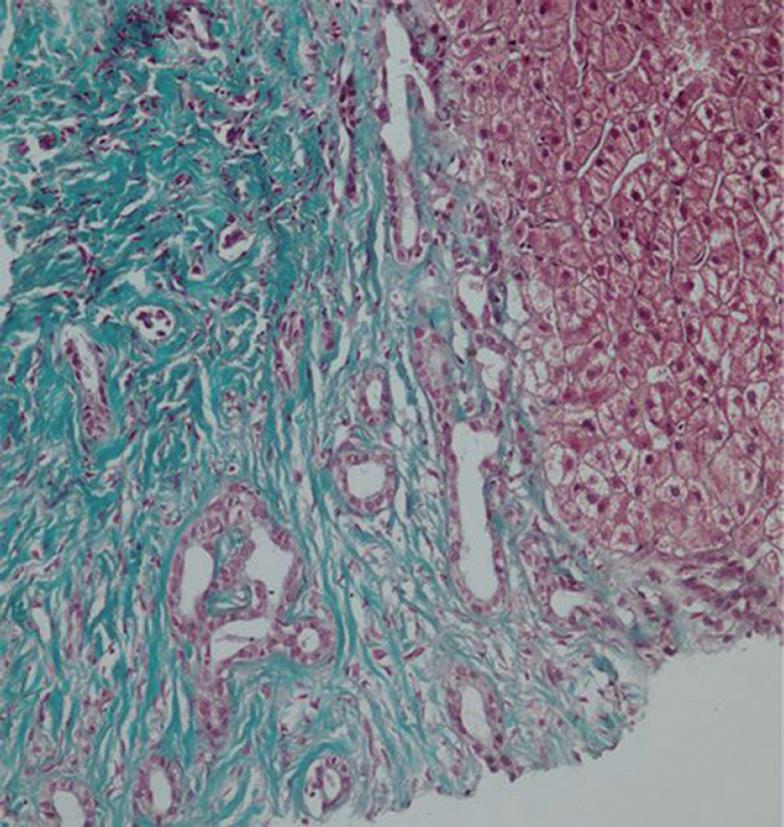
Figure 5 Liver biopsy of a patient with CHF.
The left side of the image depicts a portal area with extensive fibrosis and the presence of several bile ducts with cuboidal epithelium that have arrested at different stages of the maturation process. On the right, hepatocytes with normal morphology may be seen (× 230, trichrome stain).
- Citation: Shorbagi A, Bayraktar Y. Experience of a single center with congenital hepatic fibrosis: A review of the literature. World J Gastroenterol 2010; 16(6): 683-690
- URL: https://www.wjgnet.com/1007-9327/full/v16/i6/683.htm
- DOI: https://dx.doi.org/10.3748/wjg.v16.i6.683


