Copyright
©2010 Baishideng.
World J Gastroenterol. Feb 14, 2010; 16(6): 683-690
Published online Feb 14, 2010. doi: 10.3748/wjg.v16.i6.683
Published online Feb 14, 2010. doi: 10.3748/wjg.v16.i6.683
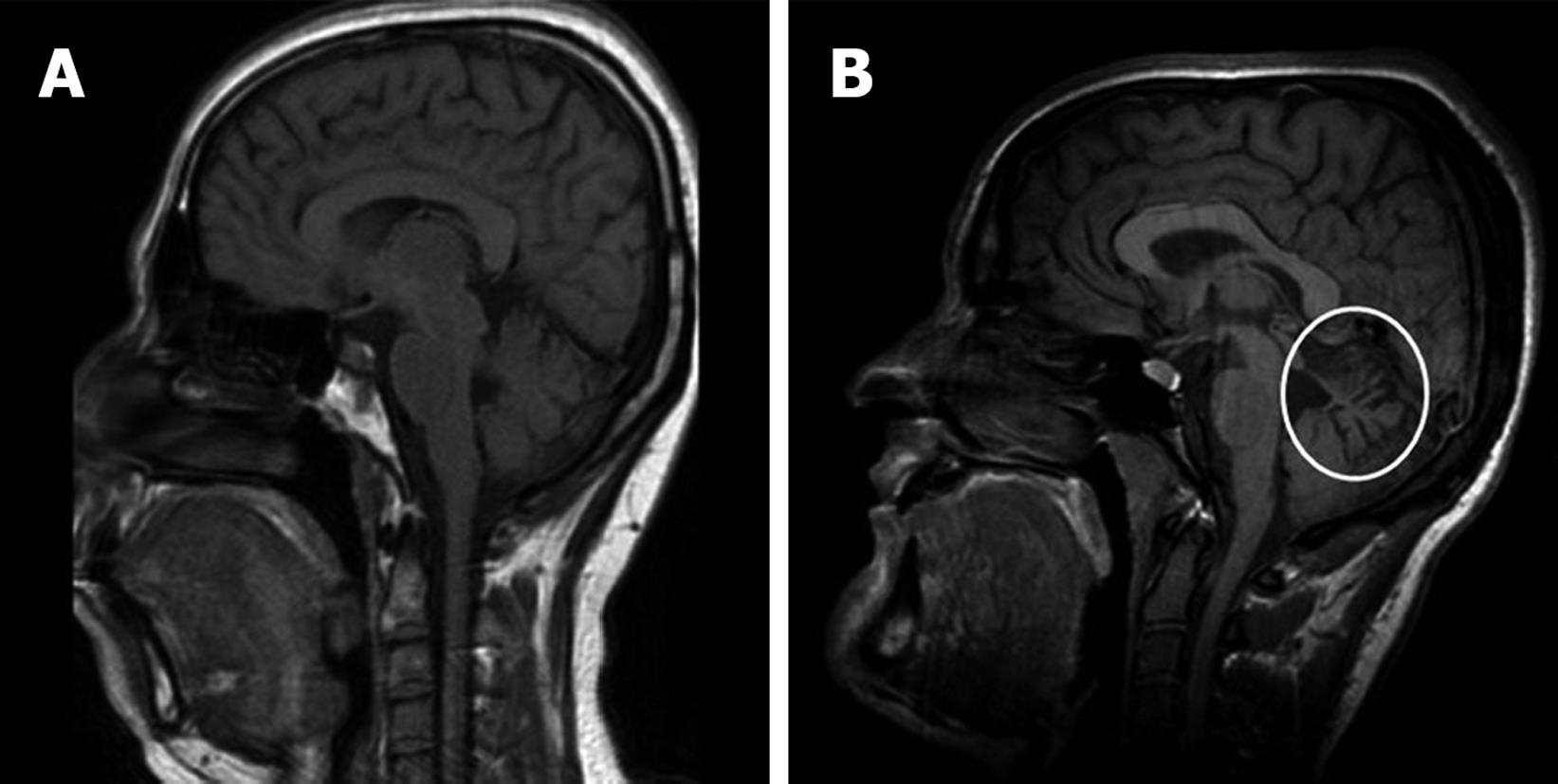
Figure 4 Brain magnetic resonance imaging (MRI) scans of two patients with congenital hepatic fibrosis.
A: A patient with Bardet-Biedl syndrome with normal findings; B: The circled area depicts cerebellar vermis atrophy manifested by more prominent folds/sulci, associated with Joubert syndrome.
- Citation: Shorbagi A, Bayraktar Y. Experience of a single center with congenital hepatic fibrosis: A review of the literature. World J Gastroenterol 2010; 16(6): 683-690
- URL: https://www.wjgnet.com/1007-9327/full/v16/i6/683.htm
- DOI: https://dx.doi.org/10.3748/wjg.v16.i6.683


