Copyright
©2010 Baishideng.
World J Gastroenterol. Feb 7, 2010; 16(5): 652-656
Published online Feb 7, 2010. doi: 10.3748/wjg.v16.i5.652
Published online Feb 7, 2010. doi: 10.3748/wjg.v16.i5.652
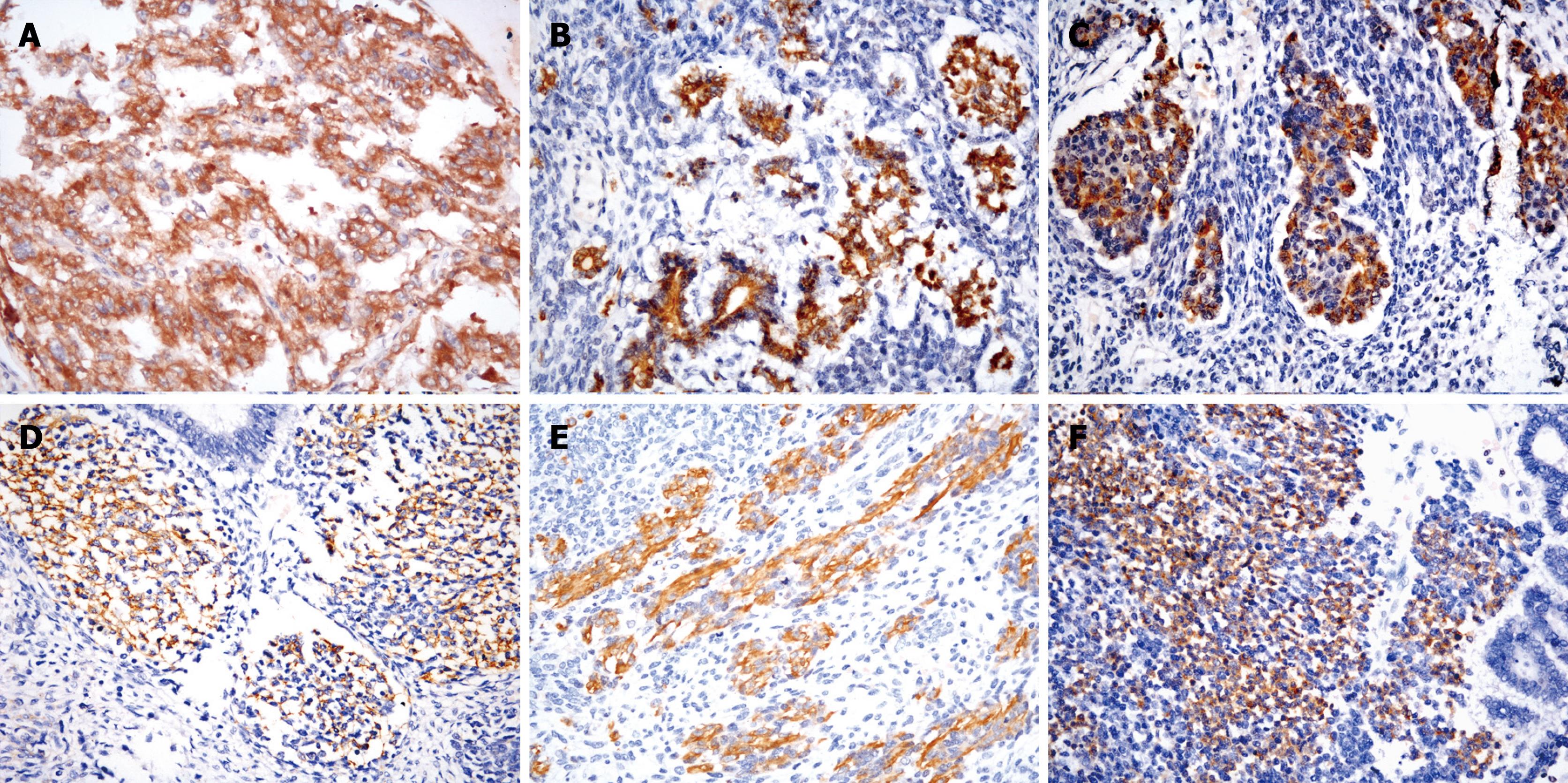
Figure 2 Immunohistochemical features of mixed GCT of the liver with sarcomatous components.
A: Strong staining for AFP in the yolk sac tumor component; B: Strong staining for AFP in acinar structures resembling pancreatic acinar tissue; C: Strong staining for α-chymotrypsin in acinar structures resembling pancreatic acinar tissue; D: CD56 positivity in primitive mesenchymal cells; E: Strong staining for desmin in rhabdomyoblastic cells in primitive mesenchymal cell components; F: Strong staining for synaptophysin in neuroblastoma tissue (EnVision Plus stain; A-F, × 400).
- Citation: Xu AM, Gong SJ, Song WH, Li XW, Pan CH, Zhu JJ, Wu MC. Primary mixed germ cell tumor of the liver with sarcomatous components. World J Gastroenterol 2010; 16(5): 652-656
- URL: https://www.wjgnet.com/1007-9327/full/v16/i5/652.htm
- DOI: https://dx.doi.org/10.3748/wjg.v16.i5.652


