Copyright
©2010 Baishideng.
World J Gastroenterol. Feb 7, 2010; 16(5): 563-570
Published online Feb 7, 2010. doi: 10.3748/wjg.v16.i5.563
Published online Feb 7, 2010. doi: 10.3748/wjg.v16.i5.563
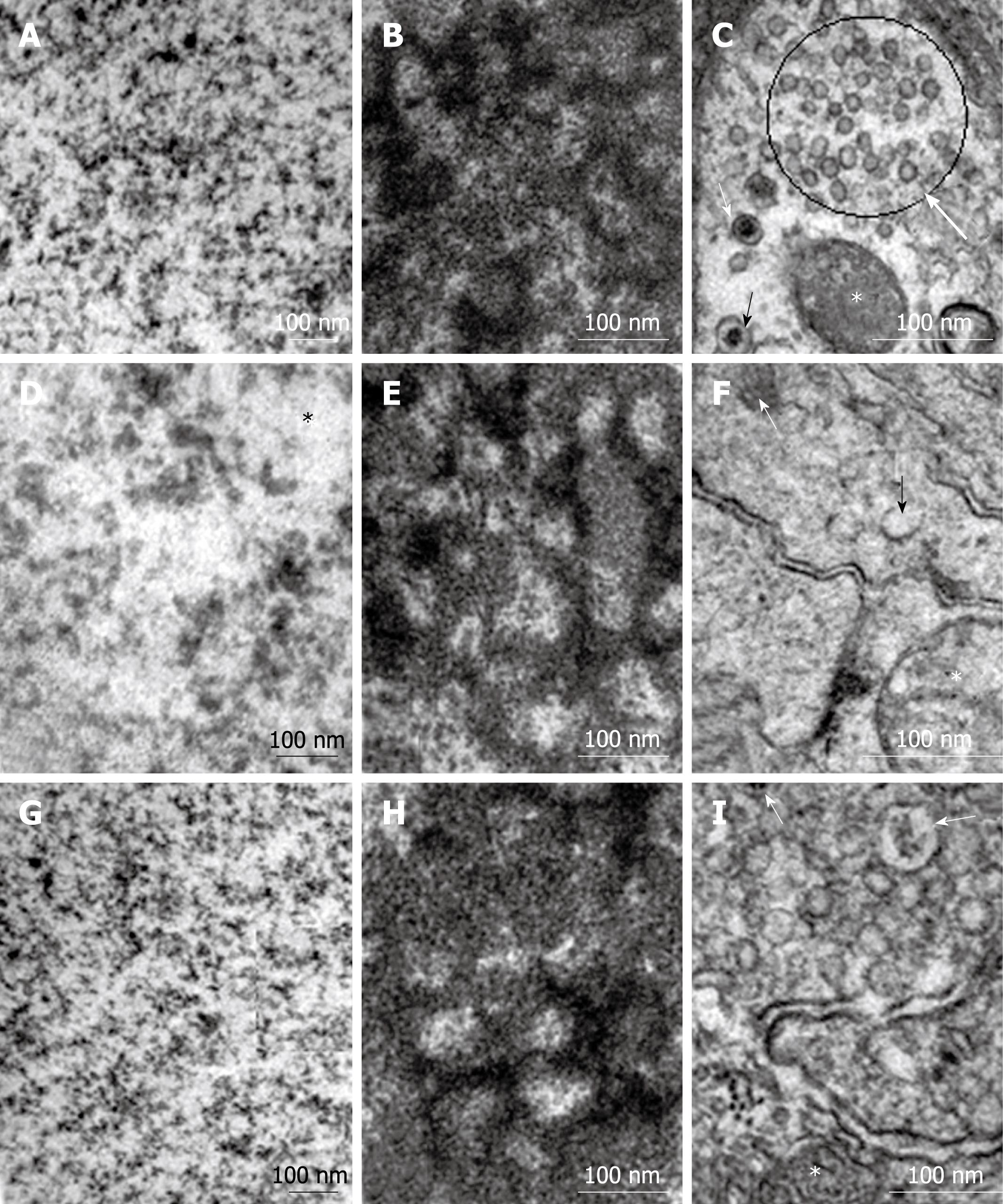
Figure 5 Electron micrographs of myenteric neurons.
A, D and G: Nuclear chromatin (*) homogeneously distributed; B, E and H: Granular and fibrillar parts of the nucleolus were similarly arranged; C, F and I: Many small vesicles (circle) were detected in neurons from all groups. Compare the intensity of large granular vesicles among the groups (arrows) (* mitochondria).
- Citation: Greggio FM, Fontes RB, Maifrino LB, Castelucci P, Souza RR, Liberti EA. Effects of perinatal protein deprivation and recovery on esophageal myenteric plexus. World J Gastroenterol 2010; 16(5): 563-570
- URL: https://www.wjgnet.com/1007-9327/full/v16/i5/563.htm
- DOI: https://dx.doi.org/10.3748/wjg.v16.i5.563


