
















©2010 Baishideng.
World J Gastroenterol. Feb 7, 2010; 16(5): 563-570
Published online Feb 7, 2010. doi: 10.3748/wjg.v16.i5.563
Published online Feb 7, 2010. doi: 10.3748/wjg.v16.i5.563
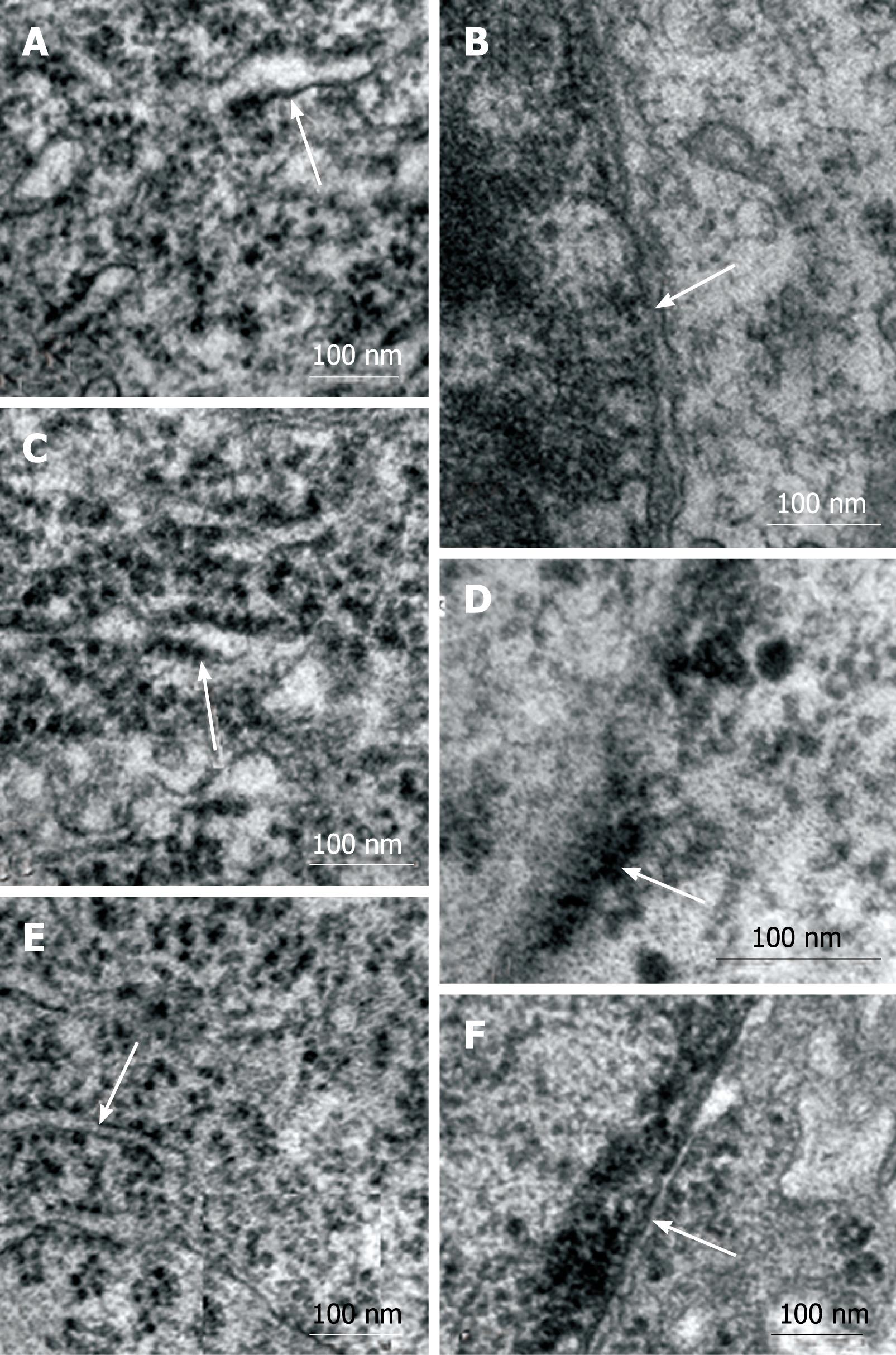
Figure 4 Electron micrographs of myenteric neurons from N42 (A and B), D42 (C and D), and R42 (E and F) groups.
A: In all groups, the granular reticulum showed that the ribosomes were aligned on the outer surface of the regularly arranged membrane (arrows). A well-defined nuclear double membrane and perinuclear cisterna were observed in neurons from N42 (B) and R42 (F) rats. Note that these structures were not delineated in the neurons of malnourished animals (D).
- Citation: Greggio FM, Fontes RB, Maifrino LB, Castelucci P, Souza RR, Liberti EA. Effects of perinatal protein deprivation and recovery on esophageal myenteric plexus. World J Gastroenterol 2010; 16(5): 563-570
- URL: https://www.wjgnet.com/1007-9327/full/v16/i5/563.htm
- DOI: https://dx.doi.org/10.3748/wjg.v16.i5.563