Copyright
©2010 Baishideng Publishing Group Co.
World J Gastroenterol. Dec 21, 2010; 16(47): 5925-5935
Published online Dec 21, 2010. doi: 10.3748/wjg.v16.i47.5925
Published online Dec 21, 2010. doi: 10.3748/wjg.v16.i47.5925
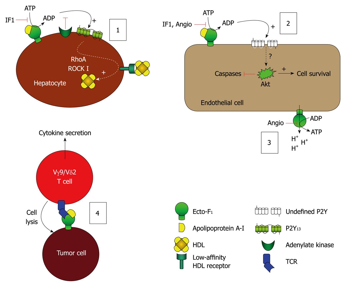
Figure 2 Model of the leading hypotheses regarding the distinct cellular events mediated by ecto-F1-ATPase.
(1) Model of F1-ATPase/P2Y13-mediated high density lipoproteins (HDL) endocytosis by hepatocytes. ApoA-I binding to ecto-F1-ATPase stimulates extracellular ATP hydrolysis, and the ADP generated selectively activates the nucleotide receptor P2Y13 and subsequent RhoA/ROCK I signaling that controls holo-HDL particle endocytosis. Conversely IF1 inhibitor which inhibits ecto-F1-ATPase activity or adenylate kinase (AK) activity which consumes ADP generated upon activation of ecto-F1-ATPase, downregulates holo-HDL particle endocytosis. Thus, HDL endocytosis depends on the balance between the AK activity that removes extracellular ADP and the ecto-F1-ATPase that synthesizes ADP. Activation of ADP synthesis by apoA-I unbalances the pathway and increases HDL endocytosis when required; (2), (3): Model of F1-ATPase-mediated endothelial cell protection. (2): Hydrolytic activity of ecto-F1-ATPase expressed on endothelial cells can be increased by apoA-I, stimulating cell proliferation. Conversely, inhibition by IF1, angiostatin increases apoptosis and blocks proliferation both at the basal level and after stimulation by apoA-I. The downstream receptors and intracellular signaling are still not completely characterized, but ADP-sensitive receptors could be involved; (3): The anti-angiogenic activity of angiostatin is attributed to its ability to inhibit ATP production by ecto-F1-ATPase, thus eliminating a significant source of energy and reducing proliferation. It is also proposed that this inhibition prevents the extrusion of protons by ecto-F1-ATPase, thus lowering intracellular pH and hampering cell survival; and (4) Model of F1-ATPase-mediated tumor cell recognition by Vγ9/Vδ2 T lymphocytes. Ecto-F1-ATPase is found at the surface of tumor cells and plays a role in the presentation of adenylated derivatives of small phosphoantigens, which are classical inducers of cell lysis by Vγ9/Vδ2 T lymphocytes. Recognition of ecto-F1-ATPase by the γ9/δ2 TCR is stimulated by apoA-I and requires a low expression of class I HLA-antigens, a situation commonly found in tumor cells.
- Citation: Vantourout P, Radojkovic C, Lichtenstein L, Pons V, Champagne E, Martinez LO. Ecto-F1-ATPase: A moonlighting protein complex and an unexpected apoA-I receptor. World J Gastroenterol 2010; 16(47): 5925-5935
- URL: https://www.wjgnet.com/1007-9327/full/v16/i47/5925.htm
- DOI: https://dx.doi.org/10.3748/wjg.v16.i47.5925


