Copyright
©2010 Baishideng Publishing Group Co.
World J Gastroenterol. Dec 14, 2010; 16(46): 5822-5829
Published online Dec 14, 2010. doi: 10.3748/wjg.v16.i46.5822
Published online Dec 14, 2010. doi: 10.3748/wjg.v16.i46.5822
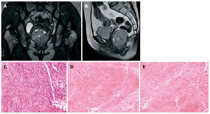
Figure 9 Retrorectal gastrointestinal stromal tumors in a 51-year-old woman.
A, B: T2-weighted magnetic resonance images show a large retrorectal mass, in which some necrosis presents hyperintensity (black arrows); C: Histopathology shows spindle cells and cytoplasmic vacuoles; D, E: On immunohistochemical studies, diffuse and strong immunoreactivity for CD117 and CD34 are seen.
- Citation: Yang BL, Gu YF, Shao WJ, Chen HJ, Sun GD, Jin HY, Zhu X. Retrorectal tumors in adults: Magnetic resonance imaging findings. World J Gastroenterol 2010; 16(46): 5822-5829
- URL: https://www.wjgnet.com/1007-9327/full/v16/i46/5822.htm
- DOI: https://dx.doi.org/10.3748/wjg.v16.i46.5822


