Copyright
©2010 Baishideng Publishing Group Co.
World J Gastroenterol. Dec 7, 2010; 16(45): 5669-5681
Published online Dec 7, 2010. doi: 10.3748/wjg.v16.i45.5669
Published online Dec 7, 2010. doi: 10.3748/wjg.v16.i45.5669
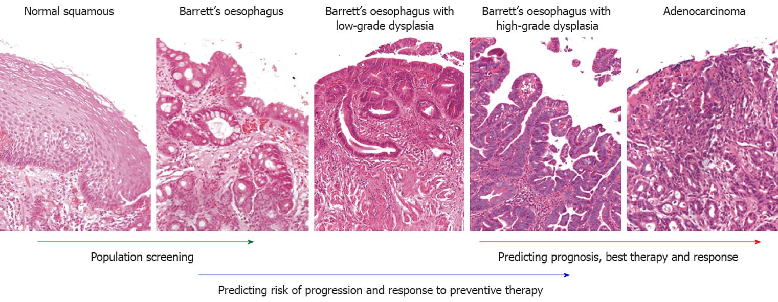
Figure 1 Transition of squamous epithelium to intestinal metaplasia, dysplasia and adenocarcinoma, with potential useful biomarkers at each stage of the disease.
The left-most panel shows normal stratified squamous epithelium. The second panel shows Barrett’s esophagus without dysplasia, with the presence of goblet cells. The third and fourth panels show Barrett’s esophagus with low-grade dysplasia and high-grade dysplasia, whereas the last panel shows adenocarcinoma.
- Citation: Ong CAJ, Lao-Sirieix P, Fitzgerald RC. Biomarkers in Barrett’s esophagus and esophageal adenocarcinoma: Predictors of progression and prognosis. World J Gastroenterol 2010; 16(45): 5669-5681
- URL: https://www.wjgnet.com/1007-9327/full/v16/i45/5669.htm
- DOI: https://dx.doi.org/10.3748/wjg.v16.i45.5669


