Copyright
copy;2010 Baishideng Publishing Group Co.
World J Gastroenterol. Oct 21, 2010; 16(39): 4998-5004
Published online Oct 21, 2010. doi: 10.3748/wjg.v16.i39.4998
Published online Oct 21, 2010. doi: 10.3748/wjg.v16.i39.4998
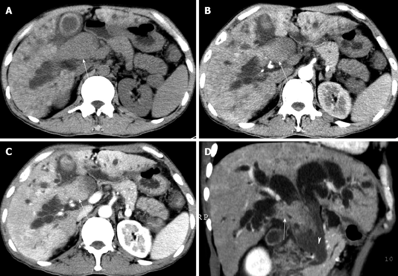
Figure 1 Axial computed tomography images of case 1 at plain scan (A), early arterial phase (B), portal phase (C), and coronal oblique plane reformation image at portal phase (D) showing irregular tumor thrombi in right hepatic, common hepatic and common bile ducts (arrows).
The tumor thrombus was mild hypo-intense on plain scan with enhancement in early arterial phase. Neither portal vein thrombus nor hepatic parenchymal mass was identified. Heterogeneously attenuated liver with lacelike fibrosis and regenerative nodules due to cirrhosis could be observed. The common bile duct was filled with hemorrhage and debris (arrowhead), which was not enhanced and mild hyper-dense in the bile.
- Citation: Long XY, Li YX, Wu W, Li L, Cao J. Diagnosis of bile duct hepatocellular carcinoma thrombus without obvious intrahepatic mass. World J Gastroenterol 2010; 16(39): 4998-5004
- URL: https://www.wjgnet.com/1007-9327/full/v16/i39/4998.htm
- DOI: https://dx.doi.org/10.3748/wjg.v16.i39.4998


