Copyright
copy;2010 Baishideng Publishing Group Co.
World J Gastroenterol. Sep 28, 2010; 16(36): 4549-4557
Published online Sep 28, 2010. doi: 10.3748/wjg.v16.i36.4549
Published online Sep 28, 2010. doi: 10.3748/wjg.v16.i36.4549
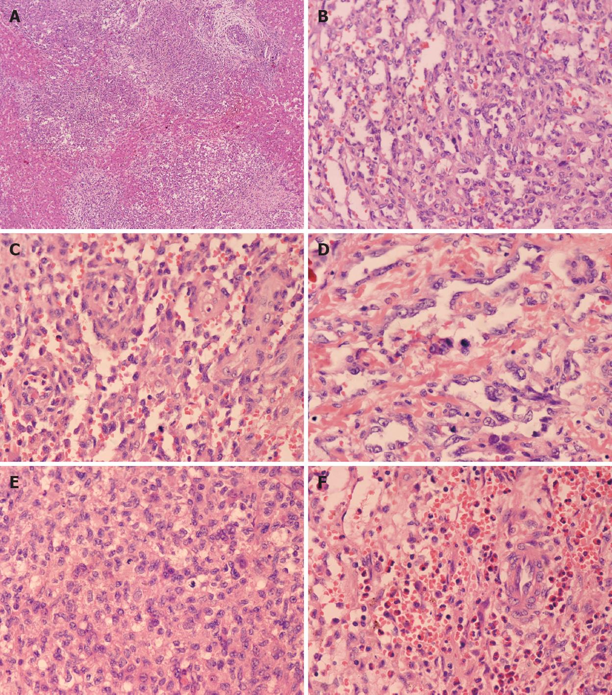
Figure 4 Histopathological features of type II lesions.
A: Multifocal nodule without encapsulation. Tumor nodules intermixed with the hepatic plate (case 5, HE, × 60); B: Anastomosing vascular structures (case 8, HE, × 300); C: A papillary structure with atypical endothelial cells (case 5, HE, × 400); D: Hyperchromatic and pleomorphic endothelial cells with mitotic cells (case 8, HE, × 400); E: Activated mitotic cells in a dense area (case 8, HE, × 400); F: Eosinophilic granulocytes were the predominant inflammatory component in one case (case 5, HE, × 400).
- Citation: Zhang Z, Chen HJ, Yang WJ, Bu H, Wei B, Long XY, Fu J, Zhang R, Ni YB, Zhang HY. Infantile hepatic hemangioendothelioma: A clinicopathologic study in a Chinese population. World J Gastroenterol 2010; 16(36): 4549-4557
- URL: https://www.wjgnet.com/1007-9327/full/v16/i36/4549.htm
- DOI: https://dx.doi.org/10.3748/wjg.v16.i36.4549


