Copyright
©2010 Baishideng Publishing Group Co.
World J Gastroenterol. Sep 14, 2010; 16(34): 4281-4290
Published online Sep 14, 2010. doi: 10.3748/wjg.v16.i34.4281
Published online Sep 14, 2010. doi: 10.3748/wjg.v16.i34.4281
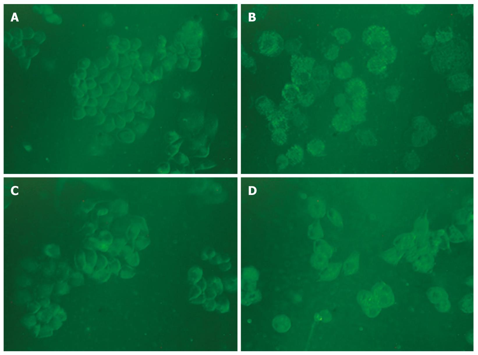
Figure 7 Monodansylcadaverine-labeled vacuoles in HepG2 cells.
Autophagic vacuoles were labeled with 0.05 mmol/L monodansylcadaverine (MDC) in phosphate-buffered saline (PBS) at 37°C for 10 min. The fluorescent density and the MDC-labeled particles in HepG2 cells were higher in matrine treatment group than in control group. The number of MDC-labeled particles in HepG2 cells was significantly lower in combined 3-MA and matrine treatment group than in single matrine treatment group (× 400 magnifications). A: Control; B: Matrine; C: 3-MA; D: 3-MA + matrine.
- Citation: Zhang JQ, Li YM, Liu T, He WT, Chen YT, Chen XH, Li X, Zhou WC, Yi JF, Ren ZJ. Antitumor effect of matrine in human hepatoma G2 cells by inducing apoptosis and autophagy. World J Gastroenterol 2010; 16(34): 4281-4290
- URL: https://www.wjgnet.com/1007-9327/full/v16/i34/4281.htm
- DOI: https://dx.doi.org/10.3748/wjg.v16.i34.4281


