Copyright
©2010 Baishideng.
World J Gastroenterol. Aug 14, 2010; 16(30): 3816-3826
Published online Aug 14, 2010. doi: 10.3748/wjg.v16.i30.3816
Published online Aug 14, 2010. doi: 10.3748/wjg.v16.i30.3816
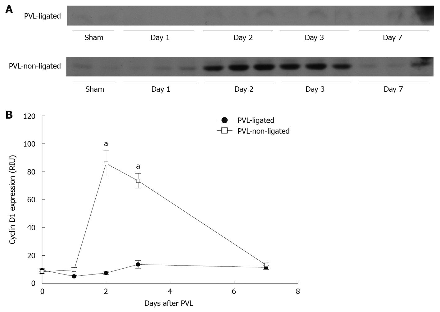
Figure 6 Liver cyclin D1 expression after portal vein ligation.
A: Liver lysates were assessed for cyclin D1 protein expression by Western blotting; B: Chemiluminescence films were quantitated by image analysis. Data are mean ± SE with n = 4 per group. aP < 0.05 vs portal vein ligation (PVL)-ligated group.
- Citation: Sakai N, Clarke CN, Schuster R, Blanchard J, Tevar AD, Edwards MJ, Lentsch AB. Portal vein ligation accelerates tumor growth in ligated, but not contralateral lobes. World J Gastroenterol 2010; 16(30): 3816-3826
- URL: https://www.wjgnet.com/1007-9327/full/v16/i30/3816.htm
- DOI: https://dx.doi.org/10.3748/wjg.v16.i30.3816


