Copyright
©2010 Baishideng.
World J Gastroenterol. Jan 21, 2010; 16(3): 384-391
Published online Jan 21, 2010. doi: 10.3748/wjg.v16.i3.384
Published online Jan 21, 2010. doi: 10.3748/wjg.v16.i3.384
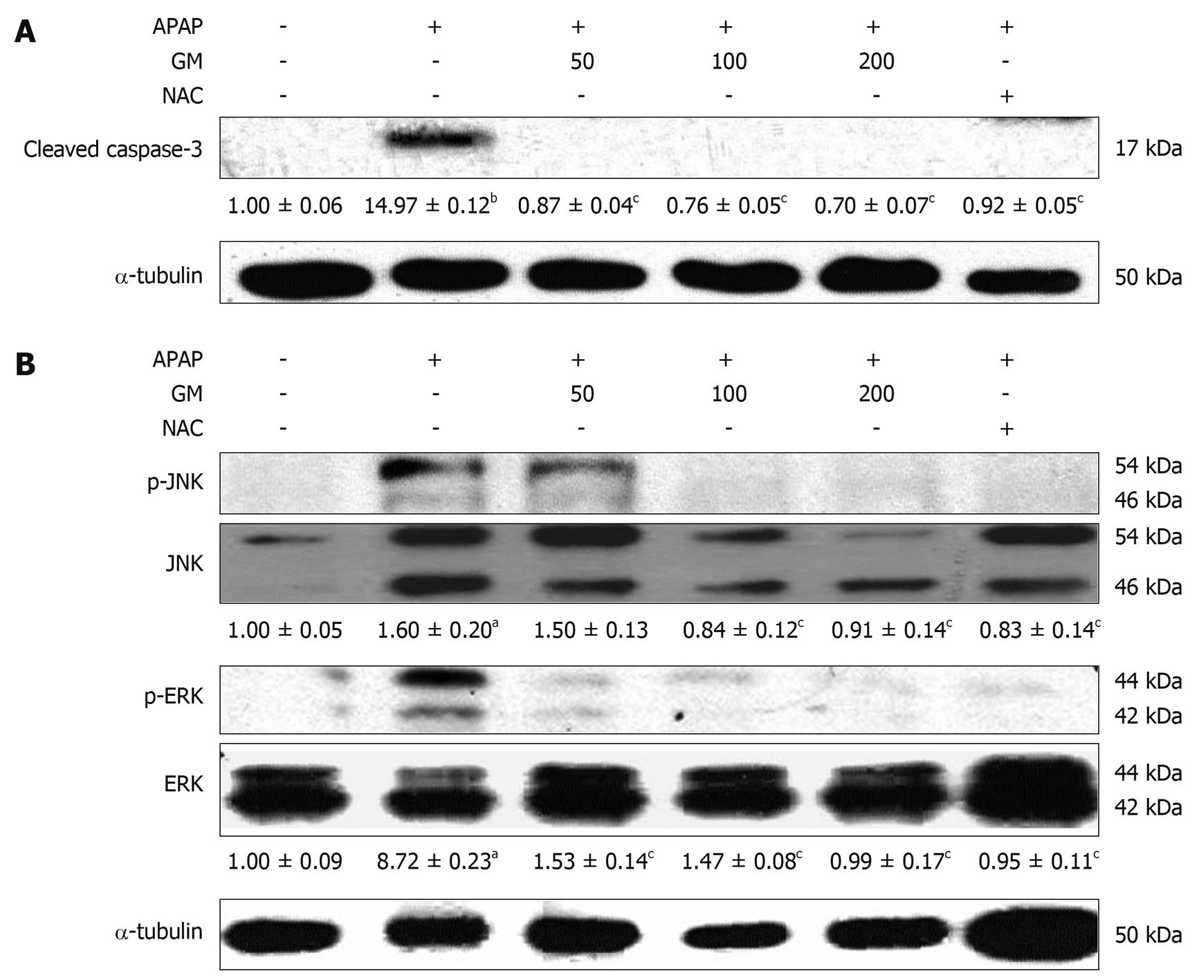
Figure 5 Western blotting analysis of caspase-3 and JNK/ERK MAPK.
GM (200, 100 or 50 mg/kg), NAC (300 mg/kg) or saline was orally administered 2 h before APAP injection. Active form of caspase-3, phospho-JNK and phospho-ERK were detected by Western blot. The active form of caspase-3 levels corresponding to each immunoreactive band were digitized and expressed as a percentage of the α-tubulin levels. Densitometric scanning data of phospho-JNK and phospho-ERK levels were expressed as the ratio of JNK or ERK, respectively. The ratio of the normal group band was set to 1.00. Data of three independent experiments are expressed as mean ± SD. aP < 0.01, bP < 0.001, significantly different when compared with the normal group. cP < 0.001 significantly different when compared with APAP alone group.
-
Citation: Wang AY, Lian LH, Jiang YZ, Wu YL, Nan JX.
Gentiana manshurica Kitagawa prevents acetaminophen-induced acute hepatic injury in micevia inhibiting JNK/ERK MAPK pathway. World J Gastroenterol 2010; 16(3): 384-391 - URL: https://www.wjgnet.com/1007-9327/full/v16/i3/384.htm
- DOI: https://dx.doi.org/10.3748/wjg.v16.i3.384


