Copyright
©2010 Baishideng.
World J Gastroenterol. Jul 28, 2010; 16(28): 3478-3483
Published online Jul 28, 2010. doi: 10.3748/wjg.v16.i28.3478
Published online Jul 28, 2010. doi: 10.3748/wjg.v16.i28.3478
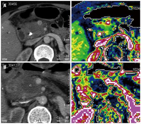
Figure 7 Pancreatic ductal adenocarcinoma before and after radiofrequency ablation.
A: Contrast-enhanced computed tomography (CT) of pancreatic head lesion that appears hypodense and vascularized at perfusion CT (right side); B: Contrast-enhanced CT of the lesion after radiofrequency ablation, which appears hypodense and avascular at perfusion CT (right side).
- Citation: D’Onofrio M, Barbi E, Girelli R, Martone E, Gallotti A, Salvia R, Martini PT, Bassi C, Pederzoli P, Mucelli RP. Radiofrequency ablation of locally advanced pancreatic adenocarcinoma: An overview. World J Gastroenterol 2010; 16(28): 3478-3483
- URL: https://www.wjgnet.com/1007-9327/full/v16/i28/3478.htm
- DOI: https://dx.doi.org/10.3748/wjg.v16.i28.3478


