Copyright
©2010 Baishideng.
World J Gastroenterol. Jul 14, 2010; 16(26): 3258-3266
Published online Jul 14, 2010. doi: 10.3748/wjg.v16.i26.3258
Published online Jul 14, 2010. doi: 10.3748/wjg.v16.i26.3258
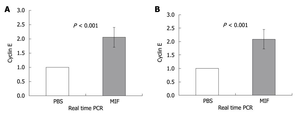
Figure 4 MIF induces cyclin E expression.
CEC were isolated from C57BL/6 (A) and NOD/SCID (B) mice that were i.p injected with MIF (400 ng) or PBS 3.5 h prior to isolation of CEC. A, B: Quantitative real time PCR was performed using primers for cyclin E and β-actin. β-actin levels were used to normalize samples for calculation of the relative expression levels of cyclin E. Results are expressed as a fold of change in cyclin E expression at stimulated cells compared to non stimulated cells, which was defined as 1. Results shown are summary of four separate experiments.
- Citation: Maharshak N, Cohen S, Lantner F, Hart G, Leng L, Bucala R, Shachar I. CD74 is a survival receptor on colon epithelial cells. World J Gastroenterol 2010; 16(26): 3258-3266
- URL: https://www.wjgnet.com/1007-9327/full/v16/i26/3258.htm
- DOI: https://dx.doi.org/10.3748/wjg.v16.i26.3258


