Copyright
©2010 Baishideng.
World J Gastroenterol. Jul 7, 2010; 16(25): 3120-3132
Published online Jul 7, 2010. doi: 10.3748/wjg.v16.i25.3120
Published online Jul 7, 2010. doi: 10.3748/wjg.v16.i25.3120
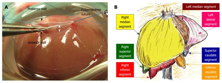
Figure 4 Hepatic segments and hepatic vein flows.
A: The hepatic vein itself has no extra-hepatic margins for suture. The most important techniques for SHIVC plasty are (1) ensuring enough margin of the wall, and (2) retention using stay sutures. In particular, sufficient margin of the SHIVC wall (white arrow) is indispensable for confirmation of optimal out-flow; B: The liver comprises 3 lobes, which are subdivided into 7 segments. In basic anatomy, the left median segment is joined with the right median segment, and an incomplete lobulation is often detected in those segments.
- Citation: Hori T, Nguyen JH, Zhao X, Ogura Y, Hata T, Yagi S, Chen F, Baine AMT, Ohashi N, Eckman CB, Herdt AR, Egawa H, Takada Y, Oike F, Sakamoto S, Kasahara M, Ogawa K, Hata K, Iida T, Yonekawa Y, Sibulesky L, Kuribayashi K, Kato T, Saito K, Wang L, Torii M, Sahara N, Kamo N, Sahara T, Yasutomi M, Uemoto S. Comprehensive and innovative techniques for liver transplantation in rats: A surgical guide. World J Gastroenterol 2010; 16(25): 3120-3132
- URL: https://www.wjgnet.com/1007-9327/full/v16/i25/3120.htm
- DOI: https://dx.doi.org/10.3748/wjg.v16.i25.3120


