Copyright
©2010 Baishideng.
World J Gastroenterol. Jul 7, 2010; 16(25): 3120-3132
Published online Jul 7, 2010. doi: 10.3748/wjg.v16.i25.3120
Published online Jul 7, 2010. doi: 10.3748/wjg.v16.i25.3120
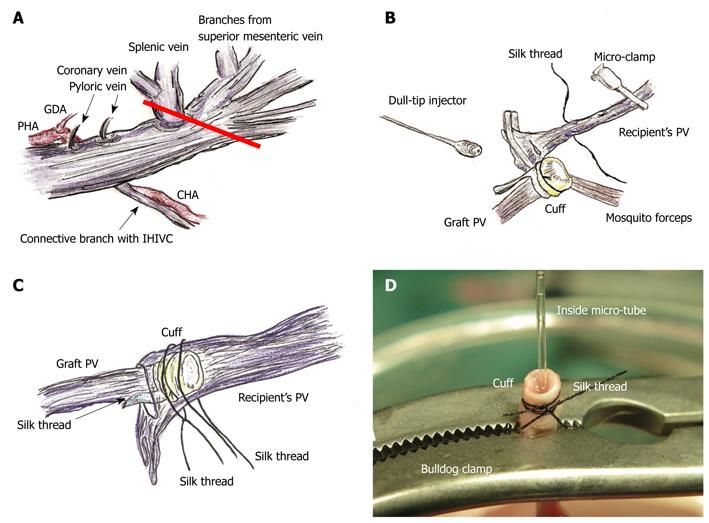
Figure 3 PV preparation and reconstruction.
A: The splenic vein and coronary vein flow into PV trunk at the left or posterior sides of the PV. The pyloric vein flows into PV trunk at the left side of PV. The posterior side of PV trunk has a branch that connects with the IHIVC. The common hepatic artery (CHA) is located at the back of PV trunk, and the proper hepatic artery (PHA) and gastroduodenal artery branches are located at the left side of PV trunk. The PV trunk and these arteries are encased together in a thin sheath. The PV trunk is cut in a branch patch-fashion using PV trunk and splenic vein in the donor operation (red line); B: Retention of recipient PV is performed using mosquito forceps. Silk thread is set behind the recipient’s PV trunk beforehand. The natural form of the PV is confirmed using saline flush. The cuff is led onto recipient’s PV. The PV is opened using the cut-down method at the point nearest the hepatic hilus, and patency of the inner side is confirmed by saline flush; C: Cuff is inserted into recipient’s PV avoiding any torsion; D: At the back table a bulldog clamp holds PV trunk, micro-tube, and cuff extension. Because of the micro-tube inside, detection of the inner side is simple. GDA: Gastroduodenal artery.
- Citation: Hori T, Nguyen JH, Zhao X, Ogura Y, Hata T, Yagi S, Chen F, Baine AMT, Ohashi N, Eckman CB, Herdt AR, Egawa H, Takada Y, Oike F, Sakamoto S, Kasahara M, Ogawa K, Hata K, Iida T, Yonekawa Y, Sibulesky L, Kuribayashi K, Kato T, Saito K, Wang L, Torii M, Sahara N, Kamo N, Sahara T, Yasutomi M, Uemoto S. Comprehensive and innovative techniques for liver transplantation in rats: A surgical guide. World J Gastroenterol 2010; 16(25): 3120-3132
- URL: https://www.wjgnet.com/1007-9327/full/v16/i25/3120.htm
- DOI: https://dx.doi.org/10.3748/wjg.v16.i25.3120


