Copyright
©2010 Baishideng.
World J Gastroenterol. Jun 21, 2010; 16(23): 2954-2958
Published online Jun 21, 2010. doi: 10.3748/wjg.v16.i23.2954
Published online Jun 21, 2010. doi: 10.3748/wjg.v16.i23.2954
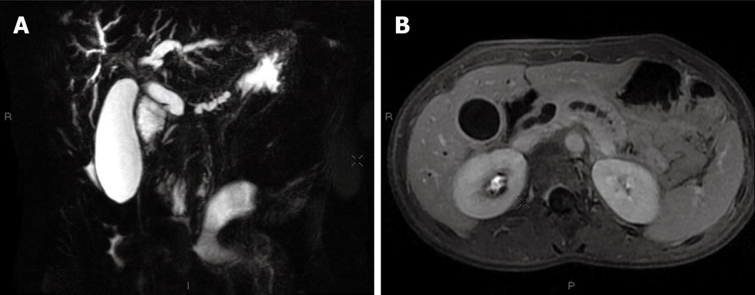
Figure 2 Initial magnetic resonance imaging (MRI) imaging.
A: T2-weighted MRI, coronal slice: bicanalar dilation and enlarged gallbladder; B: T1-weighted MRI, axial slice: main biliary duct and gallbladder enlargement with a thick wall evocative of cholangitis; the main pancreatic duct is also irregularly enlarged.
- Citation: Neuzillet C, Lepère C, Hajjam ME, Palazzo L, Fabre M, Turki H, Hammel P, Rougier P, Mitry E. Autoimmune pancreatitis with atypical imaging findings that mimicked an endocrine tumor. World J Gastroenterol 2010; 16(23): 2954-2958
- URL: https://www.wjgnet.com/1007-9327/full/v16/i23/2954.htm
- DOI: https://dx.doi.org/10.3748/wjg.v16.i23.2954


