Copyright
©2010 Baishideng.
World J Gastroenterol. Jun 14, 2010; 16(22): 2788-2792
Published online Jun 14, 2010. doi: 10.3748/wjg.v16.i22.2788
Published online Jun 14, 2010. doi: 10.3748/wjg.v16.i22.2788
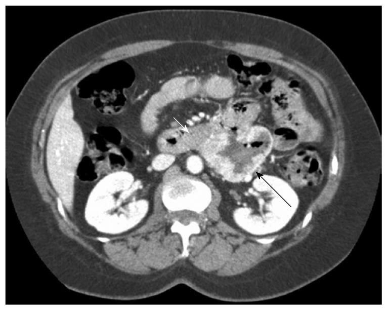
Figure 1 Gastrointestinal stromal tumor (GIST) located in the third part of the duodenum with typical computed tomography (CT) appearance.
Note the typical CT appearance of GIST with an area of necrosis (central cavitations with surrounding highly vascular tissue) (white arrow: pancreas; black arrow: GIST).
- Citation: Buchs NC, Bucher P, Gervaz P, Ostermann S, Pugin F, Morel P. Segmental duodenectomy for gastrointestinal stromal tumor of the duodenum. World J Gastroenterol 2010; 16(22): 2788-2792
- URL: https://www.wjgnet.com/1007-9327/full/v16/i22/2788.htm
- DOI: https://dx.doi.org/10.3748/wjg.v16.i22.2788


