Copyright
©2010 Baishideng.
World J Gastroenterol. Jun 14, 2010; 16(22): 2771-2779
Published online Jun 14, 2010. doi: 10.3748/wjg.v16.i22.2771
Published online Jun 14, 2010. doi: 10.3748/wjg.v16.i22.2771
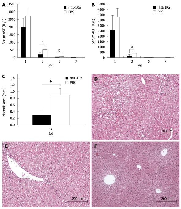
Figure 2 Acute liver injury (1 mL/kg CCl4 administration) ± rhIL-1Ra.
A: Serum aspartate aminotransferase (AST); B: Serum alanine amino transferase (ALT), with rhIL-1Ra or PBS; C: Necrotic areas. Representative findings from at least 12 mm2 tissue sections were counted for each mouse; D-F: Hematoxylin and eosin (HE) stained liver sections; D: Group received PBS at day 3 after CCl4 administration, shows necrosis with clusters of inflammatory cells around central vein (original magnification, × 100); E: Group received rhIL-1Ra at day 3 after CCl4 administration, demonstrates mostly histological recovery with only inconspicuous necrosis still remaining around central vein and very few inflammatory cells are present (original magnification, × 100); F: Normal liver histology with rhIL-1Ra treatment, which shows no difference from normal liver tissue histology. aP < 0.05, bP < 0.01.
- Citation: Zhu RZ, Xiang D, Xie C, Li JJ, Hu JJ, He HL, Yuan YS, Gao J, Han W, Yu Y. Protective effect of recombinant human IL-1Ra on CCl4-induced acute liver injury in mice. World J Gastroenterol 2010; 16(22): 2771-2779
- URL: https://www.wjgnet.com/1007-9327/full/v16/i22/2771.htm
- DOI: https://dx.doi.org/10.3748/wjg.v16.i22.2771


